

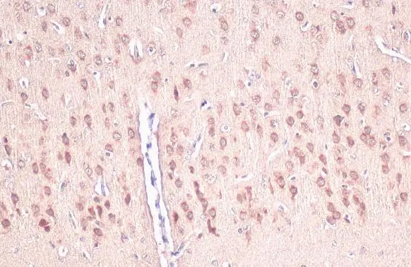
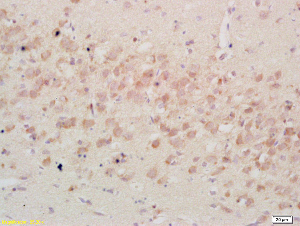
IGFBP3 antibody [N2C3] detects secreted IGFBP3 protein by immunohistochemical analysis. Sample: Paraffin-embedded rat brain. IGFBP3 stained by IGFBP3 antibody [N2C3] (GTX100454) diluted at 1:500. Antigen Retrieval: Citrate buffer, pH 6.0, 15 min
![IGFBP3 antibody [N2C3] detects IGFBP3 protein expression by immunohistochemical analysis. Sample: Frozen sectioned E13.5 Rat brain. Green: IGFBP3 protein stained by IGFBP3 antibody [N2C3] (GTX100454) diluted at 1:250. Red: beta Tubulin 3/ TUJ1, a mature neuron marker, stained by beta Tubulin 3/ TUJ1 antibody [GT11710] (GTX631836) diluted at 1:500. Blue: Fluoroshield with DAPI (GTX30920).](https://www.genetex.com/upload/website/prouct_img/normal/GTX100454/GTX100454_40604_20160921_IHC-Fr_w_23060100_135.webp "IGFBP3 antibody [N2C3] detects IGFBP3 protein expression by immunohistochemical analysis. Sample: Frozen sectioned E13.5 Rat brain. Green: IGFBP3 protein stained by IGFBP3 antibody [N2C3] (GTX100454) diluted at 1:250. Red: beta Tubulin 3/ TUJ1, a mature neuron marker, stained by beta Tubulin 3/ TUJ1 antibody [GT11710] (GTX631836) diluted at 1:500. Blue: Fluoroshield with DAPI (GTX30920).")
![U87-MG whole cell extract and conditioned medium (30 μg) were separated by 12% SDS-PAGE, and the membrane was blotted with IGFBP3 antibody [N2C3] (GTX100454) diluted at 1:1000. The HRP-conjugated anti-rabbit IgG antibody (GTX213110-01) was used to detect the primary antibody, and the signal was developed with Trident ECL plus-Enhanced.](https://www.genetex.com/upload/website/prouct_img/normal/GTX100454/GTX100454_40604_20211119_WB_Fraction_w_23060100_522.webp "U87-MG whole cell extract and conditioned medium (30 μg) were separated by 12% SDS-PAGE, and the membrane was blotted with IGFBP3 antibody [N2C3] (GTX100454) diluted at 1:1000. The HRP-conjugated anti-rabbit IgG antibody (GTX213110-01) was used to detect the primary antibody, and the signal was developed with Trident ECL plus-Enhanced.")
 was separated by 12% SDS-PAGE, and the membrane was blotted with IGFBP3 antibody (GTX100454) diluted at 1:1000. The HRP-conjugated anti-rabbit IgG antibody (GTX213110-01) was used to detect the primary antibody.")
IGFBP3 antibody [N2C3] detects secreted IGFBP3 protein by immunohistochemical analysis. Sample: Paraffin-embedded rat brain. IGFBP3 stained by IGFBP3 antibody [N2C3] (GTX100454) diluted at 1:500. Antigen Retrieval: Citrate buffer, pH 6.0, 15 min
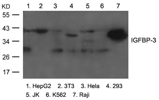
IGFBP3 antibody [N2C3]
GTX100454
ApplicationsWestern Blot, ImmunoHistoChemistry, ImmunoHistoChemistry Frozen, ImmunoHistoChemistry Paraffin
Product group Antibodies
ReactivityHuman, Mouse, Rat
TargetIGFBP3
Overview
- SupplierGeneTex
- Product NameIGFBP3 antibody [N2C3]
- Delivery Days Customer9
- Application Supplier NoteWB: 1:500-1:3000. IHC-Fr: 1:100-1:1000. *Optimal dilutions/concentrations should be determined by the researcher.Not tested in other applications.
- ApplicationsWestern Blot, ImmunoHistoChemistry, ImmunoHistoChemistry Frozen, ImmunoHistoChemistry Paraffin
- CertificationResearch Use Only
- ClonalityPolyclonal
- Concentration1.31 mg/ml
- ConjugateUnconjugated
- Gene ID3486
- Target nameIGFBP3
- Target descriptioninsulin like growth factor binding protein 3
- Target synonymsBP-53, IBP-3, IBP3, IGFBP-3, insulin-like growth factor-binding protein 3, IGF-binding protein 3, acid stable subunit of the 140 K IGF complex, binding protein 29, binding protein 53, growth hormone-dependent binding protein
- HostRabbit
- IsotypeIgG
- Protein IDP17936
- Protein NameInsulin-like growth factor-binding protein 3
- Scientific DescriptionThis gene is a member of the insulin-like growth factor binding protein (IGFBP) family and encodes a protein with an IGFBP domain and a thyroglobulin type-I domain. The protein forms a ternary complex with insulin-like growth factor acid-labile subunit (IGFALS) and either insulin-like growth factor (IGF) I or II. In this form, it circulates in the plasma, prolonging the half-life of IGFs and altering their interaction with cell surface receptors. Alternate transcriptional splice variants, encoding different isoforms, have been characterized. [provided by RefSeq]
- ReactivityHuman, Mouse, Rat
- Storage Instruction-20°C or -80°C,2°C to 8°C
- UNSPSC41116161
Datasheet
Related products
Product group Antibodies
IGFBP3 AntibodyCSB-PA003004
ApplicationsWestern Blot, ELISA, ImmunoHistoChemistry
ReactivityHuman, Mouse
TargetIGFBP3
- SizePrice

Product group Antibodies
Anti-IGFBP3 Antibody Picoband(r)A00435-2-CARRIER-FREE
ApplicationsWestern Blot, ELISA
ReactivityHuman
TargetIGFBP3
- SizePrice
Product group Antibodies
Anti-IGFBP-3 AntibodyA96572
ApplicationsWestern Blot, ELISA, ImmunoHistoChemistry
ReactivityHuman, Mouse
- SizePrice
Product group Antibodies

IGFBP3 AntibodyLS-C761132
ApplicationsWestern Blot, ImmunoHistoChemistry
ReactivityHuman, Mouse, Porcine
TargetIGFBP3
- SizePrice
Product group Antibodies

Goat anti-IGFBP3EB08005
ApplicationsWestern Blot, ELISA
ReactivityCanine, Human, Mouse, Rat
TargetIGFBP3
- SizePrice
Product group Antibodies
ApplicationsImmunoPrecipitation, Western Blot, ImmunoCytoChemistry, ImmunoHistoChemistry
ReactivityMouse
TargetIGFBP3
- SizePrice
Product group Antibodies
References
IGFBP3 Polyclonal AntibodyBS-1434R
ApplicationsImmunoFluorescence, Western Blot, ELISA, ImmunoCytoChemistry, ImmunoHistoChemistry, ImmunoHistoChemistry Frozen, ImmunoHistoChemistry Paraffin
ReactivityHuman, Mouse, Rat
TargetIGFBP3
- SizePrice
![U87-MG whole cell extract and conditioned medium (30 μg) were separated by 12% SDS-PAGE, and the membrane was blotted with IGFBP3 antibody [HL1505] (GTX636979) diluted at 1:1000. The HRP-conjugated anti-rabbit IgG antibody (GTX213110-01) was used to detect the primary antibody.](https://www.genetex.com/upload/website/prouct_img/normal/GTX636979/GTX636979_T-44676_20220513_WB_Fraction_w_23061202_477.webp)
Product group Antibodies
IGFBP3 antibody [HL1505]GTX636979
ApplicationsWestern Blot
ReactivityHuman
TargetIGFBP3
- SizePrice
Product group Antibodies
References
IGFBP3 antibody [84728.111]GTX10733
ApplicationsWestern Blot, ELISA, ImmunoHistoChemistry, ImmunoHistoChemistry Paraffin
ReactivityHuman
TargetIGFBP3
- SizePrice
Product group Antibodies
IGFBP3 antibodyGTX50853
ApplicationsWestern Blot
ReactivityHuman, Mouse
TargetIGFBP3
- SizePrice