

IHC-plus(tm) ATG9A Antibody (aa750-839)
LS-B661
ApplicationsWestern Blot, ImmunoHistoChemistry, ImmunoHistoChemistry Paraffin
Product group Antibodies
ReactivityBovine, Chicken, Human, Mouse, Primate, Rat
TargetATG9A
Overview
- SupplierLifeSpan BioSciences
- Product NameIHC-plus(tm) ATG9A Antibody (aa750-839)
- Delivery Days Customer23
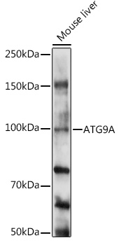
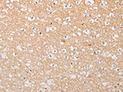
- Application Supplier NoteImmunohistochemistry: LS-B661 was validated for use in immunohistochemistry on a panel of 21 formalin-fixed, paraffin-embedded (FFPE) human tissues after heat induced antigen retrieval in pH 6.0 citrate buffer. After incubation with the primary antibody, slides were incubated with biotinylated secondary antibody, followed by alkaline phosphatase-streptavidin and chromogen. The stained slides were evaluated by a pathologist to confirm staining specificity. The optimal working concentration for LS-B661 was determined to be 5 ug/ml.. IHC, IHC-P (5 µg/ml), WB (1 - 2 µg/ml) Immunohistochemistry: LS-B661 was validated for use in immunohistochemistry on a panel of 21 formalin-fixed, paraffin-embedded (FFPE) human tissues after heat induced antigen retrieval in pH 6.0 citrate buffer. After incubation with the primary antibody, slides were incubated with biotinylated secondary antibody, followed by alkaline phosphatase-streptavidin and chromogen. The stained slides were evaluated by a pathologist to confirm staining specificity. The optimal working concentration for LS-B661 was determined to be 5 ug/ml.
- ApplicationsWestern Blot, ImmunoHistoChemistry, ImmunoHistoChemistry Paraffin
- CertificationResearch Use Only
- ClonalityPolyclonal
- Concentration1 mg/ml
- ConjugateUnconjugated
- Gene ID79065
- Target nameATG9A
- Target descriptionautophagy related 9A
- Target synonymsAPG9L1, MGD3208, mATG9, autophagy-related protein 9A, APG9 autophagy 9-like 1, APG9-like 1, ATG9 autophagy related 9 homolog A, autophagy 9-like 1 protein
- HostRabbit
- ReactivityBovine, Chicken, Human, Mouse, Primate, Rat
- UNSPSC41116161
Related products
Product group Antibodies
Anti-ATG9A Antibody144-07994
ApplicationsWestern Blot
ReactivityHuman, Mouse, Rat
TargetATG9A
- SizePrice

Product group Antibodies
Anti-ATG9A Antibody Picoband(r)A03757-2-CARRIER-FREE
ApplicationsFlow Cytometry, ImmunoFluorescence, Western Blot, ELISA, ImmunoCytoChemistry
ReactivityHuman, Mouse
TargetATG9A
- SizePrice
![ATG9A antibody detects ATG9A protein at cytoplasm by immunofluorescent analysis. Sample: HeLa cells were fixed in ice-cold MeOH for 5 min. Green: ATG9A stained by ATG9A antibody (GTX128427) diluted at 1:500. Red: alpha Tubulin, a cytoskeleton marker, stained by alpha Tubulin antibody [GT114] (GTX628802) diluted at 1:1000. Blue: Fluoroshield with DAPI (GTX30920).](https://www.genetex.com/upload/website/prouct_img/normal/GTX128427/GTX128427_44545_20220429_ICC_IF_w_23060523_217.webp)
Product group Antibodies
References
ATG9A antibodyGTX128427
ApplicationsImmunoFluorescence, ImmunoPrecipitation, Western Blot, ImmunoCytoChemistry, ImmunoHistoChemistry, ImmunoHistoChemistry Paraffin
ReactivityHuman, Mouse, Rat
TargetATG9A
- SizePrice
Product group Antibodies
ATG9A Recombinant AntibodyBSM-60696R
ApplicationsImmunoFluorescence, Western Blot, ImmunoCytoChemistry, ImmunoHistoChemistry, ImmunoHistoChemistry Frozen, ImmunoHistoChemistry Paraffin
ReactivityHuman
TargetATG9A
- SizePrice
Product group Antibodies
Anti-ATG9A AntibodyA16030
ApplicationsWestern Blot
ReactivityHuman, Mouse, Rat
- SizePrice
Product group Antibodies

ATG9A AntibodyLS-C401391
ApplicationsELISA, ImmunoHistoChemistry
ReactivityHuman, Rat
TargetATG9A
- SizePrice

Product group Antibodies
Anti-ATG9A AntibodyHPA059551
ApplicationsImmunoCytoChemistry, ImmunoHistoChemistry
ReactivityHuman
TargetATG9A
- SizePrice
Product group Antibodies
ATG9A AntibodyCSB-PA554014
ApplicationsWestern Blot, ELISA, ImmunoHistoChemistry
ReactivityHuman, Rat
TargetATG9A
- SizePrice