

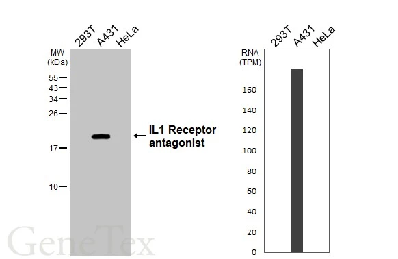
Various whole cell extracts (30 μg) were separated by 15% SDS-PAGE, and the membrane was blotted with IL1 Receptor antagonist antibody [HL3366] (GTX641186) diluted at 1:1000. The HRP-conjugated anti-rabbit IgG antibody (GTX213110-01) was used to detect the primary antibody. Corresponding RNA expression data for the same cell lines are based on Human Protein Atlas program.
![Whole cell extract (30 μg) was separated by 15% SDS-PAGE, and the membrane was blotted with IL1 Receptor antagonist antibody [HL3366] (GTX641186) diluted at 1:1000. The HRP-conjugated anti-rabbit IgG antibody (GTX213110-01) was used to detect the primary antibody.](https://www.genetex.com/upload/website/prouct_img/normal/GTX641186/GTX641186_45698_20250530_WB_D_25061003_809.webp "Whole cell extract (30 μg) was separated by 15% SDS-PAGE, and the membrane was blotted with IL1 Receptor antagonist antibody [HL3366] (GTX641186) diluted at 1:1000. The HRP-conjugated anti-rabbit IgG antibody (GTX213110-01) was used to detect the primary antibody.")
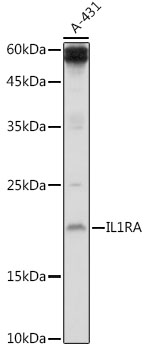
![IL1 Receptor antagonist antibody [HL3366] detects IL1 Receptor antagonist protein by immunohistochemical analysis. Sample: Paraffin-embedded human tissues. IL1 Receptor antagonist stained by IL1 Receptor antagonist antibody [HL3366] (GTX641186) diluted at 1:2500. Antigen Retrieval: Tris-EDTA buffer, pH 9.0, 15 min](https://www.genetex.com/upload/website/prouct_img/normal/GTX641186/GTX641186_45698_20250912_IHC-P_Multiple_RPKM_25091820_288.webp "IL1 Receptor antagonist antibody [HL3366] detects IL1 Receptor antagonist protein by immunohistochemical analysis. Sample: Paraffin-embedded human tissues. IL1 Receptor antagonist stained by IL1 Receptor antagonist antibody [HL3366] (GTX641186) diluted at 1:2500. Antigen Retrieval: Tris-EDTA buffer, pH 9.0, 15 min")
Various whole cell extracts (30 μg) were separated by 15% SDS-PAGE, and the membrane was blotted with IL1 Receptor antagonist antibody [HL3366] (GTX641186) diluted at 1:1000. The HRP-conjugated anti-rabbit IgG antibody (GTX213110-01) was used to detect the primary antibody. Corresponding RNA expression data for the same cell lines are based on Human Protein Atlas program.
IL1 Receptor antagonist antibody [HL3366]
GTX641186
ApplicationsWestern Blot, ImmunoHistoChemistry, ImmunoHistoChemistry Paraffin
Product group Antibodies
ReactivityHuman
TargetIL1RN
Overview
- SupplierGeneTex
- Product NameIL1 Receptor antagonist antibody [HL3366]
- Delivery Days Customer7
- ApplicationsWestern Blot, ImmunoHistoChemistry, ImmunoHistoChemistry Paraffin
- CertificationResearch Use Only
- ClonalityMonoclonal
- Clone IDHL3366
- Concentration1 mg/ml
- ConjugateUnconjugated
- Gene ID3557
- Target nameIL1RN
- Target descriptioninterleukin 1 receptor antagonist
- Target synonymsCRMO2, DIRA, ICIL-1RA, IL-1RN, IL-1ra, IL-1ra3, IL1F3, IL1RA, IRAP, MVCD4, interleukin-1 receptor antagonist protein, IL1 inhibitor, intracellular IL-1 receptor antagonist type II, intracellular interleukin-1 receptor antagonist (icIL-1ra), type II interleukin-1 receptor antagonist
- HostRabbit
- IsotypeIgG
- Protein IDP18510
- Protein NameInterleukin-1 receptor antagonist protein
- Scientific DescriptionThe protein encoded by this gene is a member of the interleukin 1 cytokine family. This protein inhibits the activities of interleukin 1, alpha (IL1A) and interleukin 1, beta (IL1B), and modulates a variety of interleukin 1 related immune and inflammatory responses. This gene and five other closely related cytokine genes form a gene cluster spanning approximately 400 kb on chromosome 2. A polymorphism of this gene is reported to be associated with increased risk of osteoporotic fractures and gastric cancer. Several alternatively spliced transcript variants encoding distinct isoforms have been reported. [provided by RefSeq, Jan 2016]
- ReactivityHuman
- Storage Instruction-20°C or -80°C,2°C to 8°C
- UNSPSC41116161
Datasheet
Related products
Product group Antibodies
IL1RN AntibodyCSB-PA06677A0RB
ApplicationsWestern Blot, ELISA
ReactivityHuman
TargetIL1RN
- SizePrice
Product group Antibodies
Il1Rn Polyclonal AntibodyCAC07246
ApplicationsWestern Blot, ELISA
TargetIL1RN
- SizePrice

Product group Antibodies
Anti-IL1RA/IL1RN Antibody Picoband(r)A00651-2-CARRIER-FREE
ApplicationsFlow Cytometry, Western Blot, ELISA, ImmunoHistoChemistry
ReactivityHuman
TargetIL1RN
- SizePrice
Product group Antibodies
Anti-IL1RN Antibody144-02088
ApplicationsImmunoFluorescence, Western Blot
ReactivityHuman, Mouse
TargetIL1RN
- SizePrice
Product group Antibodies
Anti-IL-1RA AntibodyA13858
ApplicationsImmunoFluorescence, Western Blot, ImmunoCytoChemistry
ReactivityHuman, Mouse, Rat
- SizePrice
Product group Antibodies

IL1RN AntibodyLS-C831116
ApplicationsELISA, ImmunoHistoChemistry
ReactivityHuman
TargetIL1RN
- SizePrice
Product group Antibodies
TargetIL1RN
- SizePrice

Product group Antibodies
Anti-IL1RN AntibodyHPA001482
ApplicationsWestern Blot, ImmunoHistoChemistry
ReactivityHuman
TargetIL1RN
- SizePrice
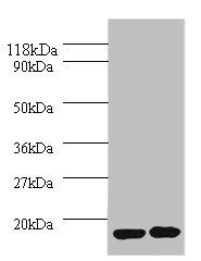
![Various whole cell extracts (30 μg) were separated by 15% SDS-PAGE, and the membrane was blotted with IL1 Receptor antagonist antibody [HL3228] (GTX640869) diluted at 1:6000. The HRP-conjugated anti-rabbit IgG antibody (GTX213110-01) was used to detect the primary antibody. Corresponding RNA expression data for the same cell lines are based on Human Protein Atlas program.](https://www.genetex.com/upload/website/prouct_img/normal/GTX640869/GTX640869_T-45509_20240823_WB_TPM_watermark_24082802_681.webp)
Product group Antibodies
ApplicationsWestern Blot
ReactivityHuman
TargetIL1RN
- SizePrice