

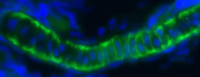
Immunofluorescent image of a pharyngeal cartilage section of a zebrafish embryo using ILK antibody [N1C1] (GTX101691) at a 1:200 dilution. ILK (Green)(This image was provided courtesy of the Schilling Lab at UC, Irvine.)
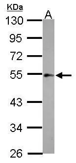
 A: zebrafish muscle 10% SDS PAGE GTX101691 diluted at 1:1000")
![Immunohistochemical analysis of paraffin-embedded zebrafish tissue, using ILK antibody [N1C1] (GTX101691) at 1:300 dilution.](https://www.genetex.com/upload/website/prouct_img/normal/GTX101691/GTX101691_39876_IHC_Z_2_22111423_383.webp "Immunohistochemical analysis of paraffin-embedded zebrafish tissue, using ILK antibody [N1C1] (GTX101691) at 1:300 dilution.")
![Immunohistochemical analysis of agarose-embedded zebrafish embryo, using ILK antibody [N1C1] (GTX101691) at 1:100 dilution. (This image was provided courtesy of the Schilling Lab at UC, Irvine.)](https://www.genetex.com/upload/website/prouct_img/normal/GTX101691/GTX101691_39876_IHC_Z_22111423_872.webp "Immunohistochemical analysis of agarose-embedded zebrafish embryo, using ILK antibody [N1C1] (GTX101691) at 1:100 dilution. (This image was provided courtesy of the Schilling Lab at UC, Irvine.)")
![Wild-type (WT) and ILK knockout (KO) HeLa cell extracts (30 μg) were separated by 10% SDS-PAGE, and the membrane was blotted with ILK antibody [N1C1] (GTX101691) diluted at 1:500. The HRP-conjugated anti-rabbit IgG antibody (GTX213110-01) was used to detect the primary antibody.](https://www.genetex.com/upload/website/prouct_img/normal/GTX101691/GTX101691_39876_20170330_WB_KO_watermark_w_23060100_182.webp "Wild-type (WT) and ILK knockout (KO) HeLa cell extracts (30 μg) were separated by 10% SDS-PAGE, and the membrane was blotted with ILK antibody [N1C1] (GTX101691) diluted at 1:500. The HRP-conjugated anti-rabbit IgG antibody (GTX213110-01) was used to detect the primary antibody.")
![Whole cell extract (30 μg) was separated by 10% SDS-PAGE, and the membrane was blotted with ILK antibody [N1C1] (GTX101691) diluted at 1:500.](https://www.genetex.com/upload/website/prouct_img/normal/GTX101691/GTX101691_39876_20160107_WB_R_w_23060100_160.webp "Whole cell extract (30 μg) was separated by 10% SDS-PAGE, and the membrane was blotted with ILK antibody [N1C1] (GTX101691) diluted at 1:500.")
![ILK antibody [N1C1] detects ILK protein at cytoplasm in mouse kidney by immunohistochemical analysis. Sample: Paraffin-embedded mouse kidney. ILK antibody [N1C1] (GTX101691) diluted at 1:500.
Antigen Retrieval: Citrate buffer, pH 6.0, 15 min](https://www.genetex.com/upload/website/prouct_img/normal/GTX101691/GTX101691_39876_20160113_IHC-P_M_w_23060100_601.webp "ILK antibody [N1C1] detects ILK protein at cytoplasm in mouse kidney by immunohistochemical analysis. Sample: Paraffin-embedded mouse kidney. ILK antibody [N1C1] (GTX101691) diluted at 1:500.
Antigen Retrieval: Citrate buffer, pH 6.0, 15 min")
![ILK antibody [N1C1] detects ILK protein by western blot analysis. A. 30 μg Neuro2A whole cell extract B. 30 μg C8D30 whole cell extract C. 30 μg NIH-3T3 whole cell extract D. 30 μg Raw 264.7 whole cell extract E. 30 μg C2Cl2 whole cell extract 10 % SDS-PAGE ILK antibody [N1C1] (GTX101691) dilution: 1:1000](https://www.genetex.com/upload/website/prouct_img/normal/GTX101691/GTX101691_39876_WB_M_w_23060100_818.webp "ILK antibody [N1C1] detects ILK protein by western blot analysis. A. 30 μg Neuro2A whole cell extract B. 30 μg C8D30 whole cell extract C. 30 μg NIH-3T3 whole cell extract D. 30 μg Raw 264.7 whole cell extract E. 30 μg C2Cl2 whole cell extract 10 % SDS-PAGE ILK antibody [N1C1] (GTX101691) dilution: 1:1000")
 A:A431(GTX27909) 7.5% SDS PAGE GTX101691 diluted at 1:500")
![ILK antibody [N1C1] immunoprecipitates ILK protein in IP experiments. IP samples: HeLa whole cell extract A. 30 μg HeLa whole cell extract B. Control with 4 μg of preimmune Rabbit IgG C. Immunoprecipitation of ILK protein by 4 μg ILK antibody [N1C1] (GTX101691) 10 % SDS-PAGE The immunoprecipitated ILK protein was detected by ILK antibody [N1C1] (GTX101691) diluted at 1:500. [EasyBlot anti-rabbit IgG (GTX221666-01) was used as a secondary reagent]](https://www.genetex.com/upload/website/prouct_img/normal/GTX101691/GTX101691_39876_IP_w_23060100_486.webp "ILK antibody [N1C1] immunoprecipitates ILK protein in IP experiments. IP samples: HeLa whole cell extract A. 30 μg HeLa whole cell extract B. Control with 4 μg of preimmune Rabbit IgG C. Immunoprecipitation of ILK protein by 4 μg ILK antibody [N1C1] (GTX101691) 10 % SDS-PAGE The immunoprecipitated ILK protein was detected by ILK antibody [N1C1] (GTX101691) diluted at 1:500. [EasyBlot anti-rabbit IgG (GTX221666-01) was used as a secondary reagent]")
Immunofluorescent image of a pharyngeal cartilage section of a zebrafish embryo using ILK antibody [N1C1] (GTX101691) at a 1:200 dilution. ILK (Green)(This image was provided courtesy of the Schilling Lab at UC, Irvine.)
ILK antibody [N1C1]
GTX101691
ApplicationsImmunoPrecipitation, Western Blot, ImmunoHistoChemistry, ImmunoHistoChemistry Paraffin
Product group Antibodies
ReactivityHuman, Mouse, Rat, Zebra Fish
TargetILK
Overview
- SupplierGeneTex
- Product NameILK antibody [N1C1]
- Delivery Days Customer9
- Application Supplier NoteWB: 1:500-1:3000. IHC-P: 1:100-1:1000. IP: 1:100-1:500. *Optimal dilutions/concentrations should be determined by the researcher.Not tested in other applications.
- ApplicationsImmunoPrecipitation, Western Blot, ImmunoHistoChemistry, ImmunoHistoChemistry Paraffin
- CertificationResearch Use Only
- ClonalityPolyclonal
- Concentration0.8 mg/ml
- ConjugateUnconjugated
- Gene ID3611
- Target nameILK
- Target descriptionintegrin linked kinase
- Target synonymsHEL-S-28, ILK-1, ILK-2, P59, p59ILK, scaffold protein ILK, 59 kDa serine/threonine-protein kinase, beta-integrin-linked kinase, epididymis secretory protein Li 28, inactive integrin-linked kinase, integrin-linked kinase-2, integrin-linked protein kinase
- HostRabbit
- IsotypeIgG
- Protein IDQ13418
- Protein NameScaffold protein ILK
- Scientific DescriptionTransduction of extracellular matrix signals through integrins influences intracellular and extracellular functions, and appears to require interaction of integrin cytoplasmic domains with cellular proteins. Integrin-linked kinase (ILK), interacts with the cytoplasmic domain of beta-1 integrin. This gene encodes a serine/threonine protein kinase with 4 ankyrin-like repeats, which associates with the cytoplasmic domain of beta integrins and acts as a proximal receptor kinase regulating integrin-mediated signal transduction. Multiple alternatively spliced transcript variants encoding the same protein have been found for this gene. [provided by RefSeq]
- ReactivityHuman, Mouse, Rat, Zebra Fish
- Storage Instruction-20°C or -80°C,2°C to 8°C
- UNSPSC41116161
Datasheet
Related products

Product group Antibodies
Anti-ILK AntibodyA96447
ApplicationsWestern Blot, ELISA, ImmunoHistoChemistry
ReactivityHuman, Mouse, Rat
- SizePrice

Product group Antibodies
Anti-Integrin linked ILK Antibody Picoband(r)A02932-2-CARRIER-FREE
ApplicationsImmunoFluorescence, Western Blot, ELISA, ImmunoCytoChemistry, ImmunoHistoChemistry
ReactivityHuman, Mouse, Rat
TargetILK
- SizePrice
Product group Antibodies
Anti-ILK Antibody144-00901
ApplicationsWestern Blot, ImmunoHistoChemistry
ReactivityHuman, Mouse, Rat
TargetILK
- SizePrice
Product group Antibodies
ILK-1 Polyclonal AntibodyBS-0317R
ApplicationsImmunoFluorescence, Western Blot, ELISA, ImmunoCytoChemistry, ImmunoHistoChemistry, ImmunoHistoChemistry Frozen, ImmunoHistoChemistry Paraffin
ReactivityBovine, Canine, Chicken, Human, Mouse, Rat
TargetILK
- SizePrice
Product group Antibodies
ILK AntibodyCSB-PA00999A0RB
ApplicationsImmunoFluorescence, Western Blot, ELISA, ImmunoHistoChemistry
ReactivityHuman, Mouse, Rat
TargetILK
- SizePrice
Product group Antibodies

Goat anti-ILKEB05120
ApplicationsWestern Blot, ELISA
ReactivityBovine, Canine, Human, Mouse, Rat
TargetILK
- SizePrice
Product group Antibodies
ILK Polyclonal AntibodyCAC13754
ApplicationsImmunoFluorescence, Western Blot, ELISA, ImmunoHistoChemistry
ReactivityMouse, Rat
TargetILK
- SizePrice
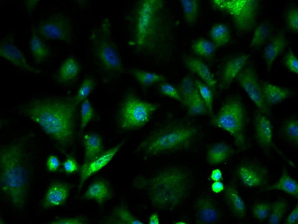
![ICC/IF analysis of PFA-fixed SH-SY-5Y cells using GTX01046 ILK antibody [SC68-04]. Green : primary antibody Blue : DAPI Permeabilization : 0.25% Triton X-100 in PBS](https://www.genetex.com/upload/website/prouct_img/normal/GTX01046/GTX01046_20200303_ICC-IF_300_w_23053121_186.webp)
Product group Antibodies
ILK antibody [SC68-04]GTX01046
ApplicationsFlow Cytometry, ImmunoFluorescence, Western Blot, ImmunoCytoChemistry, ImmunoHistoChemistry, ImmunoHistoChemistry Paraffin
ReactivityHuman, Mouse, Rat
TargetILK
- SizePrice

Product group Antibodies
Anti-ILK AntibodyHPA048437
ApplicationsWestern Blot, ImmunoCytoChemistry
ReactivityHuman
TargetILK
- SizePrice
Product group Antibodies
ILK antibody [N1C1-2]GTX107443
ApplicationsWestern Blot, ImmunoHistoChemistry, ImmunoHistoChemistry Paraffin
ReactivityHuman, Zebra Fish
TargetILK
- SizePrice