

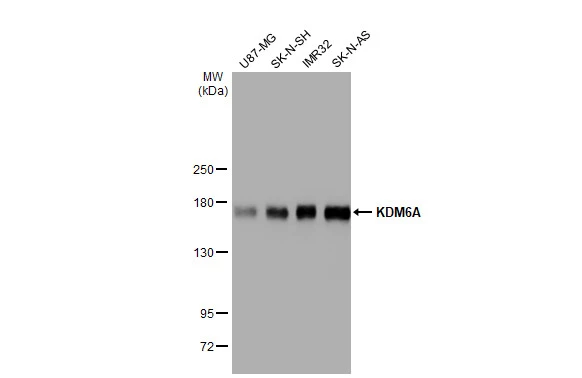
Various whole cell extracts (30 μg) were separated by 5% SDS-PAGE, and the membrane was blotted with KDM6A antibody [HL1731] (GTX637379) diluted at 1:1000. The HRP-conjugated anti-rabbit IgG antibody (GTX213110-01) was used to detect the primary antibody.
![Various whole cell extracts (30 μg) were separated by 5% SDS-PAGE, and the membrane was blotted with KDM6A antibody [HL1731] (GTX637379) diluted at 1:1000. The HRP-conjugated anti-rabbit IgG antibody (GTX213110-01) was used to detect the primary antibody. Corresponding RNA expression data for the same cell lines are based on Human Protein Atlas program.](https://www.genetex.com/upload/website/prouct_img/normal/GTX637379/GTX637379_44830_20221028_WB_TPM_watermark_22110201_391.webp "Various whole cell extracts (30 μg) were separated by 5% SDS-PAGE, and the membrane was blotted with KDM6A antibody [HL1731] (GTX637379) diluted at 1:1000. The HRP-conjugated anti-rabbit IgG antibody (GTX213110-01) was used to detect the primary antibody. Corresponding RNA expression data for the same cell lines are based on Human Protein Atlas program.")
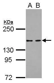
![Non-transfected (–) and transfected (+) SK-N-SH whole cell extracts (30 μg) were separated by 5% SDS-PAGE, and the membrane was blotted with KDM6A antibody [HL1731] (GTX637379) diluted at 1:1000. The HRP-conjugated anti-rabbit IgG antibody (GTX213110-01) was used to detect the primary antibody.](https://www.genetex.com/upload/website/prouct_img/normal/GTX637379/GTX637379_44830_20221028_WB_shRNA_watermark_22110201_692.webp "Non-transfected (–) and transfected (+) SK-N-SH whole cell extracts (30 μg) were separated by 5% SDS-PAGE, and the membrane was blotted with KDM6A antibody [HL1731] (GTX637379) diluted at 1:1000. The HRP-conjugated anti-rabbit IgG antibody (GTX213110-01) was used to detect the primary antibody.")
![Various whole cell extracts (30 μg) were separated by 5% SDS-PAGE, and the membrane was blotted with KDM6A antibody [HL1731] (GTX637379) diluted at 1:1000. The HRP-conjugated anti-rabbit IgG antibody (GTX213110-01) was used to detect the primary antibody.](https://www.genetex.com/upload/website/prouct_img/normal/GTX637379/GTX637379_44830_20221216_WB_D_C_22122018_545.webp "Various whole cell extracts (30 μg) were separated by 5% SDS-PAGE, and the membrane was blotted with KDM6A antibody [HL1731] (GTX637379) diluted at 1:1000. The HRP-conjugated anti-rabbit IgG antibody (GTX213110-01) was used to detect the primary antibody.")
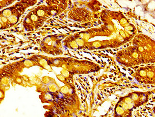
![KDM6A antibody [HL1731] detects KDM6A protein at nucleus by immunohistochemical analysis. Sample: Paraffin-embedded human breast carcinoma. KDM6A stained by KDM6A antibody [HL1731] (GTX637379) diluted at 1:100. Antigen Retrieval: Citrate buffer, pH 6.0, 15 min](https://www.genetex.com/upload/website/prouct_img/normal/GTX637379/GTX637379_44830_20221223_IHC-P_22122722_562.webp "KDM6A antibody [HL1731] detects KDM6A protein at nucleus by immunohistochemical analysis. Sample: Paraffin-embedded human breast carcinoma. KDM6A stained by KDM6A antibody [HL1731] (GTX637379) diluted at 1:100. Antigen Retrieval: Citrate buffer, pH 6.0, 15 min")
![KDM6A antibody [HL1731] detects KDM6A protein by immunofluorescent analysis. Sample: MCF-7 cells were fixed in 4% paraformaldehyde at RT for 15 min. Green: KDM6A stained by KDM6A antibody [HL1731] (GTX637379) diluted at 1:500. Red: alpha Tubulin, a cytoskeleton marker, stained by alpha Tubulin antibody [GT114] (GTX628802) diluted at 1:1000. Blue: Fluoroshield with DAPI (GTX30920). Scale bar= 10μm.](https://www.genetex.com/upload/website/prouct_img/normal/GTX637379/GTX637379_44830_20221230_ICC_IF_22122901_219.webp "KDM6A antibody [HL1731] detects KDM6A protein by immunofluorescent analysis. Sample: MCF-7 cells were fixed in 4% paraformaldehyde at RT for 15 min. Green: KDM6A stained by KDM6A antibody [HL1731] (GTX637379) diluted at 1:500. Red: alpha Tubulin, a cytoskeleton marker, stained by alpha Tubulin antibody [GT114] (GTX628802) diluted at 1:1000. Blue: Fluoroshield with DAPI (GTX30920). Scale bar= 10μm.")
![KDM6A antibody [HL1731] detects KDM6A protein by immunohistochemical analysis. Sample: Paraffin-embedded dog brain. KDM6A stained by KDM6A antibody [HL1731] (GTX637379) diluted at 1:100. Antigen Retrieval: Citrate buffer, pH 6.0, 15 min](https://www.genetex.com/upload/website/prouct_img/normal/GTX637379/GTX637379_44830_20230217_IHC-P_Dog_23030219_202.webp "KDM6A antibody [HL1731] detects KDM6A protein by immunohistochemical analysis. Sample: Paraffin-embedded dog brain. KDM6A stained by KDM6A antibody [HL1731] (GTX637379) diluted at 1:100. Antigen Retrieval: Citrate buffer, pH 6.0, 15 min")
![KDM6A antibody [HL1731] detects KDM6A protein by immunohistochemical analysis. Sample: Paraffin-embedded cat tissue. KDM6A stained by KDM6A antibody [HL1731] (GTX637379) diluted at 1:100. Antigen Retrieval: Citrate buffer, pH 6.0, 15 min](https://www.genetex.com/upload/website/prouct_img/normal/GTX637379/GTX637379_44830_20230217_IHC-P_multiple_Cat_23030219_892.webp "KDM6A antibody [HL1731] detects KDM6A protein by immunohistochemical analysis. Sample: Paraffin-embedded cat tissue. KDM6A stained by KDM6A antibody [HL1731] (GTX637379) diluted at 1:100. Antigen Retrieval: Citrate buffer, pH 6.0, 15 min")
Various whole cell extracts (30 μg) were separated by 5% SDS-PAGE, and the membrane was blotted with KDM6A antibody [HL1731] (GTX637379) diluted at 1:1000. The HRP-conjugated anti-rabbit IgG antibody (GTX213110-01) was used to detect the primary antibody.
KDM6A antibody [HL1731]
GTX637379
ApplicationsImmunoFluorescence, Western Blot, ImmunoCytoChemistry, ImmunoHistoChemistry, ImmunoHistoChemistry Paraffin
Product group Antibodies
ReactivityCanine, Feline, Human
TargetKDM6A
Overview
- SupplierGeneTex
- Product NameKDM6A antibody [HL1731]
- Delivery Days Customer9
- Application Supplier NoteWB: 1:500-1:3000. *Optimal dilutions/concentrations should be determined by the researcher.Not tested in other applications.
- ApplicationsImmunoFluorescence, Western Blot, ImmunoCytoChemistry, ImmunoHistoChemistry, ImmunoHistoChemistry Paraffin
- CertificationResearch Use Only
- ClonalityMonoclonal
- Clone IDHL1731
- Concentration1 mg/ml
- ConjugateUnconjugated
- Gene ID7403
- Target nameKDM6A
- Target descriptionlysine demethylase 6A
- Target synonymsKABUK2, UTX, bA386N14.2, lysine-specific demethylase 6A, [histone H3]-trimethyl-L-lysine(27) demethylase 6A, bA386N14.2 (ubiquitously transcribed X chromosome tetratricopeptide repeat protein (UTX)), histone demethylase UTX, lysine (K)-specific demethylase 6A, ubiquitously transcribed tetratricopeptide repeat protein X-linked, ubiquitously-transcribed TPR gene on the X chromosome
- HostRabbit
- IsotypeIgG
- Protein IDO15550
- Protein NameLysine-specific demethylase 6A
- Scientific DescriptionThis gene is located on the X chromosome and is the corresponding locus to a Y-linked gene which encodes a tetratricopeptide repeat (TPR) protein. The encoded protein of this gene contains a JmjC-domain and catalyzes the demethylation of tri/dimethylated histone H3. Multiple alternatively spliced transcript variants have been found for this gene. [provided by RefSeq, Apr 2014]
- ReactivityCanine, Feline, Human
- Storage Instruction-20°C or -80°C,2°C to 8°C
- UNSPSC41116161
Datasheet
Related products
Product group Antibodies
KDM6A AntibodyCSB-PA025774LA01HU
ApplicationsELISA, ImmunoHistoChemistry
ReactivityHuman
TargetKDM6A
- SizePrice

Product group Antibodies
Anti-UTX/KDM6A Antibody Picoband(r)A01286-2-CARRIER-FREE
ApplicationsFlow Cytometry, ImmunoFluorescence, Western Blot, ELISA, ImmunoCytoChemistry
ReactivityHuman
TargetKDM6A
- SizePrice

Product group Antibodies
Anti-KDM6A AntibodyHPA002111
ApplicationsWestern Blot, ImmunoHistoChemistry
ReactivityHuman
TargetKDM6A
- SizePrice
Product group Antibodies

KDM6A / UTX AntibodyLS-C409695
ApplicationsWestern Blot
ReactivityHuman, Mouse, Rat
TargetKDM6A
- SizePrice
Product group Antibodies
KDM6A AntibodyPACO55826
ApplicationsELISA, ImmunoHistoChemistry
ReactivityHuman
TargetKDM6A
- SizePrice
![Various whole cell extracts (50 μg) was separated by 5% SDS-PAGE, and the membrane was blotted with KDM6A antibody [HL2068] (GTX637972) diluted at 1:1000. The HRP-conjugated anti-rabbit IgG antibody (GTX213110-01) was used to detect the primary antibody.](https://www.genetex.com/upload/website/prouct_img/normal/GTX637972/GTX637972_T-44886_20221223_WB_M_22122722_423.webp)
Product group Antibodies
KDM6A antibody [HL2068]GTX637972
ApplicationsImmunoFluorescence, Western Blot, ImmunoCytoChemistry, ImmunoHistoChemistry, ImmunoHistoChemistry Paraffin
ReactivityCanine, Human, Mouse
TargetKDM6A
- SizePrice
![IHC-P analysis of human squamous cell carcinoma (SCC) from penile tissue using GTX639935 KDM6A antibody [HMV311] HistoMAX?. Squamous cell carcinoma with strong KDM6A staining of tumor cells.](https://www.genetex.com/upload/website/prouct_img/normal/GTX639935/GTX639935_20240403_IHC-P_2_24040301_816.webp)
Product group Antibodies
KDM6A antibody [HMV311] HistoMAX(tm)GTX639935
ApplicationsImmunoHistoChemistry, ImmunoHistoChemistry Paraffin
ReactivityHuman
TargetKDM6A
- SizePrice
Product group Antibodies
KDM6A antibody [C2C3], C-termGTX120873
ApplicationsWestern Blot
ReactivityHuman
TargetKDM6A
- SizePrice
![Various whole cell extracts (30 μg) were separated by 5% SDS-PAGE, and the membrane was blotted with KDM6A antibody [N2C1], Internal (GTX121246) diluted at 1:1000. The HRP-conjugated anti-rabbit IgG antibody (GTX213110-01) was used to detect the primary antibody. Corresponding RNA expression data for the same cell lines are based on Human Protein Atlas program.](https://www.genetex.com/upload/website/prouct_img/normal/GTX121246/GTX121246_42992_20221209_WB_TPM_watermark_22121123_237.webp)
Product group Antibodies
KDM6A antibody [N2C1], InternalGTX121246
ApplicationsImmunoFluorescence, ImmunoPrecipitation, Western Blot, ChIP Chromatin ImmunoPrecipitation, ImmunoCytoChemistry, ImmunoHistoChemistry, ImmunoHistoChemistry Paraffin
ReactivityHuman, Mouse
TargetKDM6A
- SizePrice