

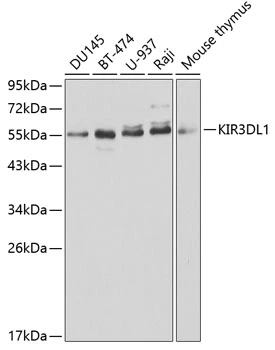
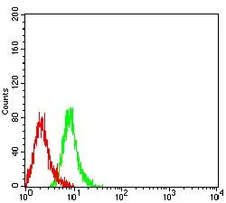
KIR3DL1 antibody [1L34]
GTX52898
ApplicationsFlow Cytometry, Neutralisation/Blocking
Product group Antibodies
ReactivityHuman
TargetKIR3DL1
Overview
- SupplierGeneTex
- Product NameKIR3DL1 antibody [1L34]
- Delivery Days Customer9
- Application Supplier NoteFCM: 1:50 - 1:100. *Optimal dilutions/concentrations should be determined by the researcher.Not tested in other applications.
- ApplicationsFlow Cytometry, Neutralisation/Blocking
- CertificationResearch Use Only
- ClonalityMonoclonal
- Clone ID1L34
- Concentration500 ug/ml
- ConjugateUnconjugated
- Gene ID3811
- Target nameKIR3DL1
- Target descriptionkiller cell immunoglobulin like receptor, three Ig domains and long cytoplasmic tail 1
- Target synonymsCD158E1, KIR, KIR2DL5B, KIR3DL1/S1, NKAT-3, NKAT3, NKB1, NKB1B, killer cell immunoglobulin-like receptor 3DL1, CD158 antigen-like family member E, HLA-BW4-specific inhibitory NK cell receptor, KIR antigen 3DL1, killer cell immunoglobulin-like receptor, three domains, long cytoplasmic tail, 1, natural killer-associated transcript 3, p70 NK receptor CL-2/CL-11, p70 killer cell inhibitory receptor, p70 natural killer cell receptor clones CL-2/CL-11
- HostMouse
- IsotypeIgG1
- Protein IDP43629
- Protein NameKiller cell immunoglobulin-like receptor 3DL1
- Scientific DescriptionKiller cell immunoglobulin-like receptors (KIRs) are transmembrane glycoproteins expressed by natural killer cells and subsets of T cells. The KIR genes are polymorphic and highly homologous and they are found in a cluster on chromosome 19q13.4 within the 1 Mb leukocyte receptor complex (LRC). The gene content of the KIR gene cluster varies among haplotypes, although several framework genes are found in all haplotypes (KIR3DL3, KIR3DP1, KIR3DL4, KIR3DL2). The KIR proteins are classified by the number of extracellular immunoglobulin domains (2D or 3D) and by whether they have a long (L) or short (S) cytoplasmic domain. KIR proteins with the long cytoplasmic domain transduce inhibitory signals upon ligand binding via an immune tyrosine-based inhibitory motif (ITIM), while KIR proteins with the short cytoplasmic domain lack the ITIM motif and instead associate with the TYRO protein tyrosine kinase binding protein to transduce activating signals. The ligands for several KIR proteins are subsets of HLA class I molecules; thus, KIR proteins are thought to play an important role in regulation of the immune response. [provided by RefSeq, Jul 2008]
- ReactivityHuman
- Storage Instruction-20°C or -80°C,2°C to 8°C
- UNSPSC41116161
Datasheet
Related products
Product group Antibodies
KIR3DL1 AntibodyCSB-PA012364LA01HU
ApplicationsImmunoFluorescence, Western Blot, ELISA, ImmunoHistoChemistry
ReactivityHuman
TargetKIR3DL1
- SizePrice
Product group Antibodies
Anti-KIR3DL1 AntibodyA30488
ApplicationsWestern Blot, ImmunoHistoChemistry
ReactivityHuman, Mouse
- SizePrice

Product group Antibodies
Anti-KIR3DL1 Antibody Picoband(r)A02187-2-CARRIER-FREE
ApplicationsWestern Blot, ELISA
ReactivityHuman
TargetKIR3DL1
- SizePrice
Product group Antibodies

KIR3DL1 AntibodyLS-C830797
ApplicationsELISA, ImmunoHistoChemistry
ReactivityHuman
TargetKIR3DL1
- SizePrice
Product group Antibodies
KIR3DL1 Polyclonal AntibodyCAC14854
ApplicationsImmunoFluorescence, Western Blot, ELISA, ImmunoHistoChemistry
TargetKIR3DL1
- SizePrice
Product group Antibodies
KIR3DL1 antibodyGTX106239
ApplicationsWestern Blot
ReactivityHuman
TargetKIR3DL1
- SizePrice
Product group Antibodies
KIR3DL1 antibody [N1C2]GTX113763
ApplicationsWestern Blot
ReactivityHuman
TargetKIR3DL1
- SizePrice
Product group Antibodies
KIR3DL1 antibody [11G25]GTX52893
ApplicationsWestern Blot
ReactivityHuman
TargetKIR3DL1
- SizePrice
Product group Antibodies
KIR3DL1 antibodyGTX54325
ApplicationsWestern Blot
ReactivityHuman, Mouse
TargetKIR3DL1
- SizePrice
Product group Antibodies
KIR3DL1 antibody [6D9A4]GTX00648
ApplicationsFlow Cytometry, Western Blot, ELISA
ReactivityHuman, Rat
TargetKIR3DL1
- SizePrice