

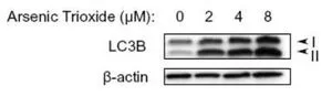
WB analysis of arsenic trioxide treated U87-MG cell lysate using GTX82986 LC3B antibody.


WB analysis of arsenic trioxide treated U87-MG cell lysate using GTX82986 LC3B antibody.
LC3B antibody
GTX82986
ApplicationsElectron Microscopy, ImmunoFluorescence, Western Blot, ImmunoCytoChemistry, ImmunoHistoChemistry, ImmunoHistoChemistry Frozen, ImmunoHistoChemistry Paraffin
Product group Antibodies
ReactivityBovine, Canine, Human, Mouse, Porcine, Primate, Rat, Zebra Fish
TargetMAP1LC3B
Overview
- SupplierGeneTex
- Product NameLC3B antibody
- Delivery Days Customer9
- Application Supplier NoteWB: 1:1000. ICC/IF: 0.1 - 2 microg/ml. IHC-P: 1:200 - 1:400. IHC-Fr: 1:400. *Optimal dilutions/concentrations should be determined by the researcher.Not tested in other applications.
- ApplicationsElectron Microscopy, ImmunoFluorescence, Western Blot, ImmunoCytoChemistry, ImmunoHistoChemistry, ImmunoHistoChemistry Frozen, ImmunoHistoChemistry Paraffin
- CertificationResearch Use Only
- ClonalityPolyclonal
- Concentration1 mg/ml
- ConjugateUnconjugated
- Gene ID81631
- Target nameMAP1LC3B
- Target descriptionmicrotubule associated protein 1 light chain 3 beta
- Target synonymsATG8F, LC3B, MAP1A/1BLC3, MAP1LC3B-a, microtubule-associated protein 1 light chain 3 beta, MAP1 light chain 3-like protein 2, MAP1A/MAP1B LC3 B, MAP1A/MAP1B light chain 3 B, autophagy-related ubiquitin-like modifier LC3 B, microtubule-associated proteins 1A/1B light chain 3B
- HostRabbit
- IsotypeIgG
- Protein IDQ9GZQ8
- Protein NameMicrotubule-associated protein 1 light chain 3 beta
- Scientific DescriptionThe product of this gene is a subunit of neuronal microtubule-associated MAP1A and MAP1B proteins, which are involved in microtubule assembly and important for neurogenesis. Studies on the rat homolog implicate a role for this gene in autophagy, a process that involves the bulk degradation of cytoplasmic component. [provided by RefSeq]
- ReactivityBovine, Canine, Human, Mouse, Porcine, Primate, Rat, Zebra Fish
- Storage Instruction-20°C or -80°C,2°C to 8°C
- UNSPSC12352203
References
- Varga G, Schleifenbaum S, Koenig U, et al. Phagocytic cell death leads to enhanced release of pro-inflammatory S100A12 in familial Mediterranean fever. Mol Cell Pediatr. 2023,10(1):19. doi: 10.1186/s40348-023-00173-3Read this paper
- Mostafa DK, Nayel OA, Abdulmalek S, et al. Modulation of autophagy, apoptosis and oxidative stress: a clue for repurposing metformin in photoaging. Inflammopharmacology. 2022,30(6):2521-2535. doi: 10.1007/s10787-022-01041-8Read this paper
- Pan X, Taherzadeh M, Bose P, et al. Glucosamine amends CNS pathology in mucopolysaccharidosis IIIC mouse expressing misfolded HGSNAT. J Exp Med. 2022,219(8). doi: 10.1084/jem.20211860Read this paper
- Tsai CH, Lii CK, Wang TS, et al. Docosahexaenoic acid promotes the formation of autophagosomes in MCF-7 breast cancer cells through oxidative stress-induced growth inhibitor 1 mediated activation of AMPK/mTOR pathway. Food Chem Toxicol. 2021,154:112318. doi: 10.1016/j.fct.2021.112318Read this paper
- Mohapatra S, Chakraborty T, Shimizu S, et al. Estrogen and estrogen receptors chauffeur the sex-biased autophagic action in liver. Cell Death Differ. 2020,27(11):3117-3130. doi: 10.1038/s41418-020-0567-3Read this paper
- Koenig U, Robenek H, Barresi C, et al. Cell death induced autophagy contributes to terminal differentiation of skin and skin appendages. Autophagy. 2020,16(5):932-945. doi: 10.1080/15548627.2019.1646552Read this paper
- Sukseree S, Chen YT, Laggner M, et al. Tyrosinase-Cre-Mediated Deletion of the Autophagy Gene Atg7 Leads to Accumulation of the RPE65 Variant M450 in the Retinal Pigment Epithelium of C57BL/6 Mice. PLoS One. 2016,11(8):e0161640. doi: 10.1371/journal.pone.0161640Read this paper
- Koenig U, Fobker M, Lengauer B, et al. Autophagy facilitates secretion and protects against degeneration of the Harderian gland. Autophagy. 2015,11(2):298-313. doi: 10.4161/15548627.2014.978221Read this paper
- Rossiter H, König U, Barresi C, et al. Epidermal keratinocytes form a functional skin barrier in the absence of Atg7 dependent autophagy. J Dermatol Sci. 2013,71(1):67-75. doi: 10.1016/j.jdermsci.2013.04.015Read this paper
- Zhao Y, Zhang CF, Rossiter H, et al. Autophagy is induced by UVA and promotes removal of oxidized phospholipids and protein aggregates in epidermal keratinocytes. J Invest Dermatol. 2013,133(6):1629-37. doi: 10.1038/jid.2013.26Read this paper
Datasheet
Related products
Product group Antibodies

Cleaved LC3B AntibodyABX029981
ApplicationsImmunoFluorescence, ELISA, ImmunoCytoChemistry
- SizePrice
Product group Antibodies
Anti-MAP1LC3B Antibody144-11282
ApplicationsImmunoFluorescence, Western Blot, ImmunoHistoChemistry
ReactivityHuman, Mouse, Porcine, Rat
TargetMAP1LC3B
- SizePrice
Product group Antibodies
References
LC3B antibody [N1C3]GTX116080
ApplicationsFlow Cytometry, ImmunoFluorescence, Western Blot, ImmunoCytoChemistry, ImmunoHistoChemistry, ImmunoHistoChemistry Paraffin
ReactivityHuman, Mouse, Rat
TargetMAP1LC3B
- SizePrice
![ICC/IF analysis of PFA-fixed HeLa cells with/without Chloroquine (50 μM, 37oC, 20 hrs) treatment using GTX00949 LC3B antibody [GT1187]. Right : untreated HeLa cells Left : HeLa cells with 50 μM Chloroquine (20 hrs, 37oC) treatment Orange : Primary antibody Blue : DAPI Dilution : 1:100](https://www.genetex.com/upload/website/prouct_img/normal/GTX00949/GTX00949_20200327_ICC-IF_43_w_23053121_449.webp)
Product group Antibodies
LC3B antibody [GT1187]GTX00949
ApplicationsImmunoFluorescence, Western Blot, ImmunoCytoChemistry, ImmunoHistoChemistry, ImmunoHistoChemistry Paraffin
ReactivityHuman, Mouse, Rat
TargetMAP1LC3B
- SizePrice
![FACS analysis of HeLa cells using GTX60594 LC3B antibody [5H12]. Green : LC3B Red : negative control](https://www.genetex.com/upload/website/prouct_img/normal/GTX60594/GTX60594_20170912_FACS_w_23061123_292.webp)
Product group Antibodies
LC3B antibody [5H12]GTX60594
ApplicationsFlow Cytometry, Western Blot, ELISA
ReactivityHuman
TargetMAP1LC3B
- SizePrice
![Untreated (–) and treated (+) HeLa whole cell extracts (50 μg) were separated by 15% SDS-PAGE, and the membrane was blotted with LC3B antibody [GT3612] (GTX632501) diluted at 1:500.](https://www.genetex.com/upload/website/prouct_img/normal/GTX632501/GTX632501_42261_20151231_WB_chloroquine_w_23061202_146.webp)
Product group Antibodies
References
LC3B antibody [GT3612]GTX632501
ApplicationsImmunoFluorescence, Western Blot, ImmunoCytoChemistry, ImmunoHistoChemistry, ImmunoHistoChemistry Paraffin
ReactivityHuman
TargetMAP1LC3B
- SizePrice
Product group Antibodies
Map1Lc3B Polyclonal AntibodyCAC09906
ApplicationsELISA, ImmunoHistoChemistry
TargetMAP1LC3B
- SizePrice
Product group Antibodies
References
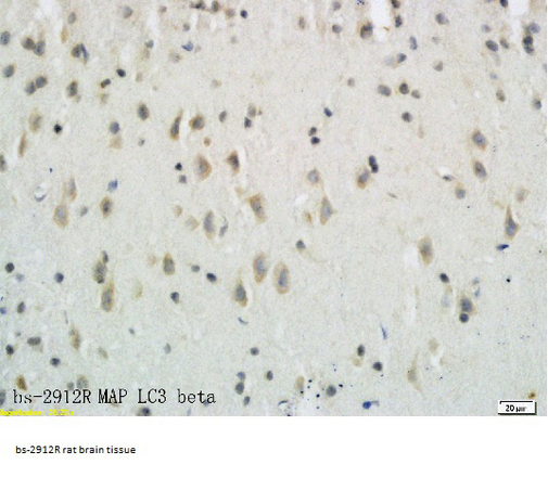
LC3B Polyclonal AntibodyBS-2912R
ApplicationsImmunoFluorescence, Western Blot, ELISA, ImmunoCytoChemistry, ImmunoHistoChemistry, ImmunoHistoChemistry Frozen, ImmunoHistoChemistry Paraffin
ReactivityBovine, Canine, Chicken, Equine, Human, Mouse, Porcine, Rabbit, Rat, Zebra Fish
TargetMAP1LC3B
- SizePrice
Product group Antibodies
ReactivityHuman
TargetMAP1LC3B
- SizePrice