

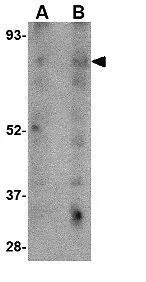
WB analysis of human skeletal muscle tissue lysate using GTX85031 LIMP II antibody. Working concentration : (A) 1 and (B) 2 μg/ml

WB analysis of human skeletal muscle tissue lysate using GTX85031 LIMP II antibody. Working concentration : (A) 1 and (B) 2 μg/ml
LIMP II antibody
GTX85031
ApplicationsWestern Blot, ELISA, ImmunoHistoChemistry, ImmunoHistoChemistry Paraffin
Product group Antibodies
ReactivityHuman, Mouse
TargetSCARB2
Overview
- SupplierGeneTex
- Product NameLIMP II antibody
- Delivery Days Customer9
- Application Supplier NoteWB: 1 - 2 microg/mL. IHC-P: 10 microg/mL. *Optimal dilutions/concentrations should be determined by the researcher.Not tested in other applications.
- ApplicationsWestern Blot, ELISA, ImmunoHistoChemistry, ImmunoHistoChemistry Paraffin
- CertificationResearch Use Only
- ClonalityPolyclonal
- Concentration1 mg/ml
- ConjugateUnconjugated
- Gene ID950
- Target nameSCARB2
- Target descriptionscavenger receptor class B member 2
- Target synonymsAMRF, CD36L2, EPM4, HLGP85, LGP85, LIMP-2, LIMPII, SR-BII, lysosome membrane protein 2, 85 kDa lysosomal membrane sialoglycoprotein, 85 kDa lysosomal sialoglycoprotein scavenger receptor class B, member 2, CD36 antigen (collagen type I receptor, thrombospondin receptor)-like 2 (lysosomal integral membrane protein II), CD36 antigen-like 2, LIMP II, lysosome membrane protein II
- HostRabbit
- IsotypeIgG
- Protein IDQ14108
- Protein NameLysosome membrane protein 2
- Scientific DescriptionThe lysosomal integral membrane protein 2 (LIMP2) is a heavily glycosylated type III transmembrane protein, the majority of which exists in the lumen of the lysosome and a cytoplasmic domain of approximately 20 amino acids. A deficiency of LIMP2 in mice causes uretic pelvic junction obstruction, deafness, and peripheral neuropathy associated with impaired vesicular trafficking and distribution of apically expressed proteins. More recently, LIMP2 was shown to act as a receptor to bind b-glucocerebrosidase, the enzyme defective in Gaucher disease, a lysosomal storage disorder. LIMP2-deficient mice showed missorted as well as secreted b-glucocerebrosidase, suggesting that LIMP2 also functions as the mannose-6-phosphate-independent trafficking receptor. Despite its predicted molecular weight, LIMP2 runs at approximately 80 - 85 kDa in SDS-PAGE.
- ReactivityHuman, Mouse
- Storage Instruction-20°C or -80°C,2°C to 8°C
- UNSPSC12352203
Datasheet
Related products

Product group Antibodies
Anti-SCARB2 Antibody144-12723
ApplicationsWestern Blot
ReactivityHuman, Mouse, Rat
TargetSCARB2
- SizePrice

Product group Antibodies
Anti-Scavenging Receptor SRB2/SCARB2 Antibody Picoband(r)A05090-1-CARRIER-FREE
ApplicationsFlow Cytometry, ImmunoFluorescence, Western Blot, ELISA, ImmunoHistoChemistry
ReactivityHuman
TargetSCARB2
- SizePrice
Product group Antibodies
Scarb2 Polyclonal AntibodyCAC07916
ApplicationsImmunoFluorescence, Western Blot, ELISA, ImmunoHistoChemistry
ReactivityMouse
TargetSCARB2
- SizePrice
Product group Antibodies
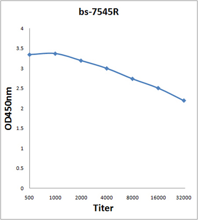
Scavenger Receptor BII AntibodyBS-7545R
ApplicationsImmunoFluorescence, Western Blot, ELISA, ImmunoCytoChemistry, ImmunoHistoChemistry, ImmunoHistoChemistry Frozen, ImmunoHistoChemistry Paraffin
ReactivityBovine, Canine, Equine, Human, Mouse, Porcine, Rabbit, Rat
TargetSCARB2
- SizePrice
Product group Antibodies

LIMP2 AntibodyABX430331
ApplicationsWestern Blot, ELISA, ImmunoHistoChemistry
- SizePrice
Product group Antibodies
SCARB2 AntibodyCSB-PA619859LA01HU
ApplicationsImmunoFluorescence, Western Blot, ELISA, ImmunoHistoChemistry
ReactivityHuman, Mouse
TargetSCARB2
- SizePrice
Product group Antibodies

ApplicationsWestern Blot, ELISA, ImmunoHistoChemistry
ReactivityHuman, Mouse
TargetSCARB2
- SizePrice
Product group Antibodies
TargetSCARB2
- SizePrice