

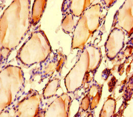
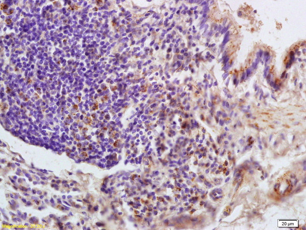
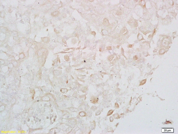
Immunohistochemistry of paraffin-embedded human thyroid tissue using CSB-PA013032LA01HU at dilution of 1:100
")
Immunohistochemistry of paraffin-embedded human thyroid tissue using CSB-PA013032LA01HU at dilution of 1:100
LONP1 Antibody
CSB-PA013032LA01HU
ApplicationsImmunoFluorescence, ELISA, ImmunoHistoChemistry
Product group Antibodies
ReactivityHuman
TargetLONP1
Overview
- SupplierCusabio
- Product NameLONP1 Antibody
- Delivery Days Customer20
- ApplicationsImmunoFluorescence, ELISA, ImmunoHistoChemistry
- CertificationResearch Use Only
- ClonalityPolyclonal
- ConjugateUnconjugated
- Gene ID9361
- Target nameLONP1
- Target descriptionlon peptidase 1, mitochondrial
- Target synonymsCODASS, LON, LONP, LonHS, PIM1, PRSS15, hLON, lon protease homolog, mitochondrial, hLON ATP-dependent protease, mitochondrial ATP-dependent protease Lon, mitochondrial lon protease-like protein, serine protease 15
- HostRabbit
- IsotypeIgG
- Protein IDP36776
- Protein NameLon protease homolog, mitochondrial
- Scientific DescriptionATP-dependent serine protease that mediates the selective degradation of misfolded, unassembled or oxidatively damaged polypeptides as well as certain short-lived regulatory proteins in the mitochondrial matrix. May also have a chaperone function in the assembly of inner membrane protein complexes. Participates in the regulation of mitochondrial gene expression and in the maintenance of the integrity of the mitochondrial genome. Binds to mitochondrial promoters and RNA in a single-stranded, site-specific, and strand-specific manner. May regulate mitochondrial DNA replication and/or gene expression using site-specific, single-stranded DNA binding to target the degradation of regulatory proteins binding to adjacent sites in mitochondrial promoters. Endogenous substrates include mitochondrial steroidogenic acute regulatory (StAR) protein.
- ReactivityHuman
- Storage Instruction-20°C or -80°C
- UNSPSC41116161
Related products
Product group Antibodies
Anti-LONP1 AntibodyA38188
ApplicationsWestern Blot, ImmunoHistoChemistry
ReactivityHuman, Mouse
- SizePrice

Product group Antibodies
Anti-LONP1/Lon Antibody Picoband(r)A03808-2-CARRIER-FREE
ApplicationsFlow Cytometry, ImmunoFluorescence, Western Blot, ELISA, ImmunoCytoChemistry, ImmunoHistoChemistry
ReactivityHuman, Mouse, Rat
TargetLONP1
- SizePrice
Product group Antibodies

LONP1 / LON AntibodyLS-C667905
ApplicationsWestern Blot
ReactivityHuman
TargetLONP1
- SizePrice
Product group Antibodies
References
LONP1 Polyclonal AntibodyBS-4245R
ApplicationsImmunoFluorescence, Western Blot, ELISA, ImmunoCytoChemistry, ImmunoHistoChemistry, ImmunoHistoChemistry Frozen, ImmunoHistoChemistry Paraffin
ReactivityBovine, Canine, Chicken, Equine, Human, Mouse, Porcine, Rat
TargetLONP1
- SizePrice

Product group Antibodies
Anti-LONP1 AntibodyHPA002192
ApplicationsWestern Blot, ImmunoCytoChemistry, ImmunoHistoChemistry
ReactivityHuman, Mouse, Rat
TargetLONP1
- SizePrice
Product group Antibodies
References
LONP1 antibodyGTX51596
ApplicationsWestern Blot, ImmunoHistoChemistry, ImmunoHistoChemistry Paraffin
ReactivityHuman, Mouse, Rat
TargetLONP1
- SizePrice