

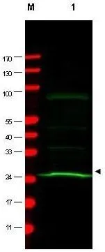
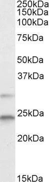
Western blot using GeneTex Mab anti-MAD2L1 antibody (GTX10691) shows detection of a band at ~24 kDa (arrowhead) corresponding to MAD2L1 present in a HeLa whole cell lysate (lane 1). Approximately 75 μg of lysate was separated by 4-20% TG SDS-PAGE. After blocking, the membrane was probed overnight at 4oC with the primary antibody diluted to 1:200. The membrane was washed and reacted with a 1:5,000 dilution of IRDye?800 conjugated Sh-a-Mouse IgG [H&L] for 45 min at room temperature (800 nm channel, green). Molecular weight estimation was made by comparison to prestained MW markers in lane M (700 nm channel, red). IRDye?800 fluorescence image was captured using the OdysseyR Infrared Imaging System developed by LI-COR. IRDye is a trademark of LI-COR, Inc.
![WB analysis of HeLa whole cell lysate using GTX10691 MAD2L1 antibody [17D10]. Loading : 75 μg Dilution : 1:200](https://www.genetex.com/upload/website/prouct_img/normal/GTX10691/GTX10691_20240423_WB_9_24042320_323.webp "WB analysis of HeLa whole cell lysate using GTX10691 MAD2L1 antibody [17D10]. Loading : 75 μg Dilution : 1:200")
Western blot using GeneTex Mab anti-MAD2L1 antibody (GTX10691) shows detection of a band at ~24 kDa (arrowhead) corresponding to MAD2L1 present in a HeLa whole cell lysate (lane 1). Approximately 75 μg of lysate was separated by 4-20% TG SDS-PAGE. After blocking, the membrane was probed overnight at 4oC with the primary antibody diluted to 1:200. The membrane was washed and reacted with a 1:5,000 dilution of IRDye?800 conjugated Sh-a-Mouse IgG [H&L] for 45 min at room temperature (800 nm channel, green). Molecular weight estimation was made by comparison to prestained MW markers in lane M (700 nm channel, red). IRDye?800 fluorescence image was captured using the OdysseyR Infrared Imaging System developed by LI-COR. IRDye is a trademark of LI-COR, Inc.
MAD2L1 antibody [17D10]
GTX10691
ApplicationsImmunoPrecipitation, Western Blot, ELISA
Product group Antibodies
ReactivityHuman
TargetMAD2L1
Overview
- SupplierGeneTex
- Product NameMAD2L1 antibody [17D10]
- Delivery Days Customer9
- Application Supplier NoteWB: 1:200-1:2000. IP: 1:100. ELISA: 1:5000-1:20000. *Optimal dilutions/concentrations should be determined by the researcher.Not tested in other applications.
- ApplicationsImmunoPrecipitation, Western Blot, ELISA
- CertificationResearch Use Only
- ClonalityMonoclonal
- Clone ID17D10
- Concentration1 mg/ml
- ConjugateUnconjugated
- Gene ID4085
- Target nameMAD2L1
- Target descriptionmitotic arrest deficient 2 like 1
- Target synonymsHSMAD2, MAD2, mitotic spindle assembly checkpoint protein MAD2A, MAD2 (mitotic arrest deficient, yeast, homolog)-like 1, MAD2 mitotic arrest deficient-like 1, MAD2-like protein 1, mitotic arrest deficient 2-like protein 1, mitotic arrest deficient, yeast, homolog-like 1
- HostMouse
- IsotypeIgG1
- Protein IDQ13257
- Protein NameMitotic spindle assembly checkpoint protein MAD2A
- Scientific DescriptionMAD2L1 (also called mitotic spindle assembly checkpoint protein, MAD2A, MAD2-like 1 and HsMAD2) is a component of the mitotic spindle assembly checkpoint monitors the process of kinetochore-spindle attachment and delays the onset of anaphase when this process is not complete. MAD2L1 inhibits the activity of the anaphase-promoting complex by sequestering CDC20 until all chromosomes are aligned at the metaphase plate. MAD2L1 is related to the MAD2L2 gene located on chromosome 1. This protein has a nuclear localization.
- ReactivityHuman
- Storage Instruction-20°C or -80°C,2°C to 8°C
- UNSPSC41116161
Datasheet
Related products
Product group Antibodies
Anti-MAD2L1 AntibodyA100502
ApplicationsWestern Blot, ELISA
ReactivityHuman
- SizePrice
Product group Antibodies
MAD2L1 Recombinant AntibodyBSM-54219R
ApplicationsImmunoFluorescence, Western Blot, ImmunoCytoChemistry, ImmunoHistoChemistry, ImmunoHistoChemistry Paraffin
ReactivityHuman
TargetMAD2L1
- SizePrice
Product group Antibodies
MAD2L1 AntibodyCSB-PA003184
ApplicationsWestern Blot, ELISA
ReactivityHuman
TargetMAD2L1
- SizePrice
Product group Antibodies

Goat anti-MAD2L1EB05612
ApplicationsWestern Blot, ELISA
ReactivityBovine, Canine, Human, Mouse, Porcine, Rat
TargetMAD2L1
- SizePrice
Product group Antibodies
Mad2L1 Polyclonal AntibodyCAC10698
ApplicationsWestern Blot, ELISA, ImmunoHistoChemistry
TargetMAD2L1
- SizePrice
Product group Antibodies
MAD2L1 antibody, C-termGTX13615
ApplicationsWestern Blot
ReactivityHuman
TargetMAD2L1
- SizePrice
Product group Antibodies

MAD2L1 / MAD2 Antibody (aa2-205, FITC)LS-C371381
ApplicationsELISA
ReactivityHuman
TargetMAD2L1
- SizePrice
![ICC/IF analysis of COS7 cells transiently transfected with MAD2L1 plasmid using GTX84169 MAD2L1 antibody [4D2].](https://www.genetex.com/upload/website/prouct_img/normal/GTX84169/GTX84169_976_ICCIF_w_23061420_252.webp)
Product group Antibodies
MAD2L1 antibody [4D2]GTX84169
ApplicationsImmunoFluorescence, Western Blot, ImmunoCytoChemistry
ReactivityHuman
TargetMAD2L1
- SizePrice
Product group Antibodies
Anti-MAD2L1 AntibodyHPA003348
ApplicationsWestern Blot, ImmunoCytoChemistry, ImmunoHistoChemistry
ReactivityHuman
TargetMAD2L1
- SizePrice
Product group Antibodies
MAD2L1 antibody [C2C3], C-termGTX104680
ApplicationsImmunoFluorescence, ImmunoPrecipitation, Western Blot, ImmunoCytoChemistry, ImmunoHistoChemistry, ImmunoHistoChemistry Paraffin
ReactivityHuman, Rat
TargetMAD2L1
- SizePrice