

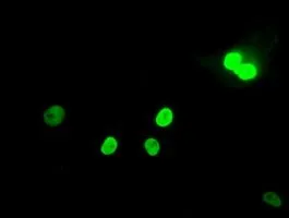
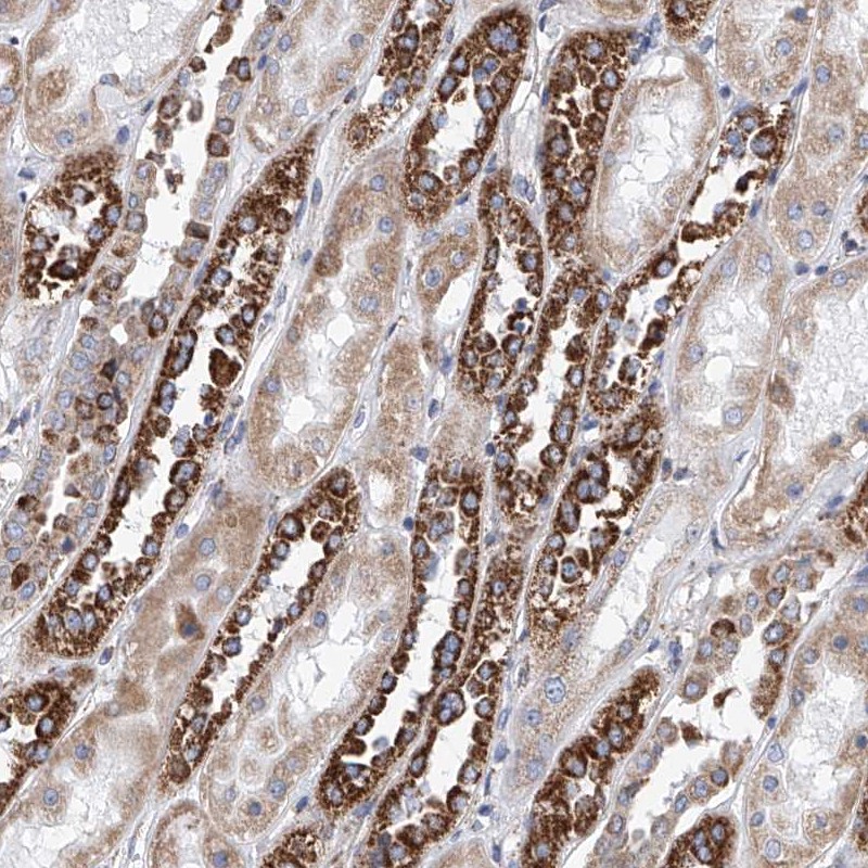
ICC/IF analysis of COS7 cells transiently transfected with MAD2L1 plasmid using GTX84169 MAD2L1 antibody [4D2].
![WB analysis of HEK293T cells transfected with MAD2L1 plasmid (Right) or empty vector (Left) for 48 hrs using GTX84169 MAD2L1 antibody [4D2]. Loading : 5 ug per lane](https://www.genetex.com/upload/website/prouct_img/normal/GTX84169/GTX84169_4173_WB_w_23061420_826.webp "WB analysis of HEK293T cells transfected with MAD2L1 plasmid (Right) or empty vector (Left) for 48 hrs using GTX84169 MAD2L1 antibody [4D2]. Loading : 5 ug per lane")
ICC/IF analysis of COS7 cells transiently transfected with MAD2L1 plasmid using GTX84169 MAD2L1 antibody [4D2].
MAD2L1 antibody [4D2]
GTX84169
ApplicationsImmunoFluorescence, Western Blot, ImmunoCytoChemistry
Product group Antibodies
ReactivityHuman
TargetMAD2L1
Overview
- SupplierGeneTex
- Product NameMAD2L1 antibody [4D2]
- Delivery Days Customer9
- Application Supplier NoteWB: 1:2000. ICC/IF: 1:100. *Optimal dilutions/concentrations should be determined by the researcher.Not tested in other applications.
- ApplicationsImmunoFluorescence, Western Blot, ImmunoCytoChemistry
- CertificationResearch Use Only
- ClonalityMonoclonal
- Clone ID4D2
- Concentration1 mg/ml
- ConjugateUnconjugated
- Gene ID4085
- Target nameMAD2L1
- Target descriptionmitotic arrest deficient 2 like 1
- Target synonymsHSMAD2, MAD2, mitotic spindle assembly checkpoint protein MAD2A, MAD2 (mitotic arrest deficient, yeast, homolog)-like 1, MAD2 mitotic arrest deficient-like 1, MAD2-like protein 1, mitotic arrest deficient 2-like protein 1, mitotic arrest deficient, yeast, homolog-like 1
- HostMouse
- IsotypeIgG2a
- Protein IDQ13257
- Protein NameMitotic spindle assembly checkpoint protein MAD2A
- Scientific DescriptionComponent of the spindle-assembly checkpoint that prevents the onset of anaphase until all chromosomes are properly aligned at the metaphase plate. Required for the execution of the mitotic checkpoint which monitors the process of kinetochore-spindle attachment and inhibits the activity of the anaphase promoting complex by sequestering CDC20 until all chromosomes are aligned at the metaphase plate.
- ReactivityHuman
- Storage Instruction-20°C or -80°C,2°C to 8°C
- UNSPSC41116161
Datasheet
Related products
Product group Antibodies
Anti-MAD2L1 AntibodyA100502
ApplicationsWestern Blot, ELISA
ReactivityHuman
- SizePrice
Product group Antibodies
MAD2L1 Recombinant AntibodyBSM-54219R
ApplicationsImmunoFluorescence, Western Blot, ImmunoCytoChemistry, ImmunoHistoChemistry, ImmunoHistoChemistry Paraffin
ReactivityHuman
TargetMAD2L1
- SizePrice
Product group Antibodies
MAD2L1 AntibodyCSB-PA003184
ApplicationsWestern Blot, ELISA
ReactivityHuman
TargetMAD2L1
- SizePrice
Product group Antibodies

Goat anti-MAD2L1EB05612
ApplicationsWestern Blot, ELISA
ReactivityBovine, Canine, Human, Mouse, Porcine, Rat
TargetMAD2L1
- SizePrice
Product group Antibodies
Mad2L1 Polyclonal AntibodyCAC10698
ApplicationsWestern Blot, ELISA, ImmunoHistoChemistry
TargetMAD2L1
- SizePrice
Product group Antibodies
MAD2L1 antibody, C-termGTX13615
ApplicationsWestern Blot
ReactivityHuman
TargetMAD2L1
- SizePrice
Product group Antibodies

MAD2L1 / MAD2 Antibody (aa2-205, FITC)LS-C371381
ApplicationsELISA
ReactivityHuman
TargetMAD2L1
- SizePrice
Product group Antibodies
Anti-MAD2L1 AntibodyHPA003348
ApplicationsWestern Blot, ImmunoCytoChemistry, ImmunoHistoChemistry
ReactivityHuman
TargetMAD2L1
- SizePrice
Product group Antibodies
MAD2L1 antibody [C2C3], C-termGTX104680
ApplicationsImmunoFluorescence, ImmunoPrecipitation, Western Blot, ImmunoCytoChemistry, ImmunoHistoChemistry, ImmunoHistoChemistry Paraffin
ReactivityHuman, Rat
TargetMAD2L1
- SizePrice
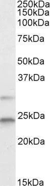
![Western blot using GeneTex Mab anti-MAD2L1 antibody (GTX10691) shows detection of a band at ~24 kDa (arrowhead) corresponding to MAD2L1 present in a HeLa whole cell lysate (lane 1). Approximately 75 μg of lysate was separated by 4-20% TG SDS-PAGE. After blocking, the membrane was probed overnight at 4oC with the primary antibody diluted to 1:200. The membrane was washed and reacted with a 1:5,000 dilution of IRDye?800 conjugated Sh-a-Mouse IgG [H&L] for 45 min at room temperature (800 nm channel, green). Molecular weight estimation was made by comparison to prestained MW markers in lane M (700 nm channel, red). IRDye?800 fluorescence image was captured using the OdysseyR Infrared Imaging System developed by LI-COR. IRDye is a trademark of LI-COR, Inc.](https://www.genetex.com/upload/website/prouct_img/normal/GTX10691/GTX10691_20160330_WB_w_23060120_790.webp)
Product group Antibodies
MAD2L1 antibody [17D10]GTX10691
ApplicationsImmunoPrecipitation, Western Blot, ELISA
ReactivityHuman
TargetMAD2L1
- SizePrice