

MADCAM1 Antibody (YA2744)
HY-P82999
TargetMADCAM1
Product group Antibodies
Overview
- SupplierMedChem Express
- Product NameMADCAM1 Antibody (YA2744)
- Delivery Days Customer5
- CertificationResearch Use Only
- ClonalityMonoclonal
- Gene ID8174
- Target nameMADCAM1
- Target descriptionmucosal vascular addressin cell adhesion molecule 1
- Target synonymshMAdCAM-1; MACAM1; MAdCAM-1; mucosal addressin cell adhesion molecule 1
- HostRabbit
- IsotypeIgG
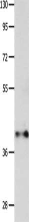
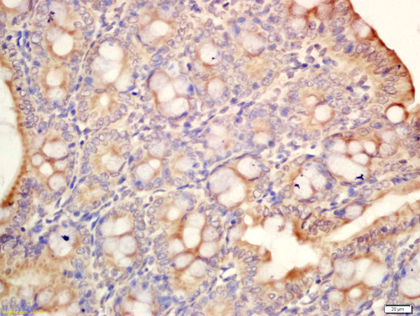
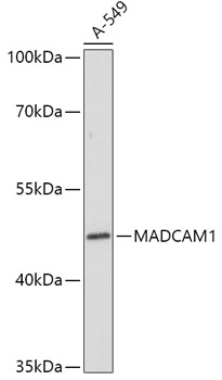
- Scientific DescriptionMADCAM1 Antibody (YA2744) is a non-conjugated IgG antibody, targeting MADCAM1, with a predicted molecular weight of 40 kDa (observed band size: 60 kDa). MADCAM1 Antibody (YA2744) can be used for WB experiment in human background.
- UNSPSC12352203
Related products
Product group Antibodies
MADCAM1 AntibodyCSB-PA295662
ApplicationsWestern Blot, ELISA
TargetMADCAM1
- SizePrice
Product group Antibodies
MAdCAM1 Polyclonal AntibodyBS-11179R
ApplicationsFlow Cytometry, ImmunoFluorescence, Western Blot, ELISA, ImmunoCytoChemistry, ImmunoHistoChemistry, ImmunoHistoChemistry Frozen, ImmunoHistoChemistry Paraffin
TargetMADCAM1
- SizePrice
Product group Antibodies
Madcam1 Polyclonal AntibodyCAC10448
ApplicationsImmunoFluorescence, Western Blot, ELISA
TargetMADCAM1
- SizePrice
Product group Antibodies
MAdCAM-1 antibody [314G8]GTX39768
ApplicationsFlow Cytometry, ELISA, ImmunoHistoChemistry, ImmunoHistoChemistry Frozen
TargetMADCAM1
- SizePrice

Product group Antibodies
Anti-MADCAM1 AntibodyHPA077998
ApplicationsImmunoHistoChemistry
TargetMADCAM1
- SizePrice
Product group Antibodies

MAdCAM-1 AntibodyLS-C408632
ApplicationsWestern Blot, ImmunoHistoChemistry, ImmunoHistoChemistry Paraffin
TargetMADCAM1
- SizePrice

Product group Antibodies
Anti-MAdCAM1 Antibody Picoband(r)A04227-1-CARRIER-FREE
ApplicationsWestern Blot
TargetMADCAM1
- SizePrice