

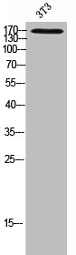
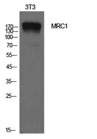
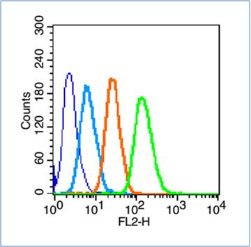
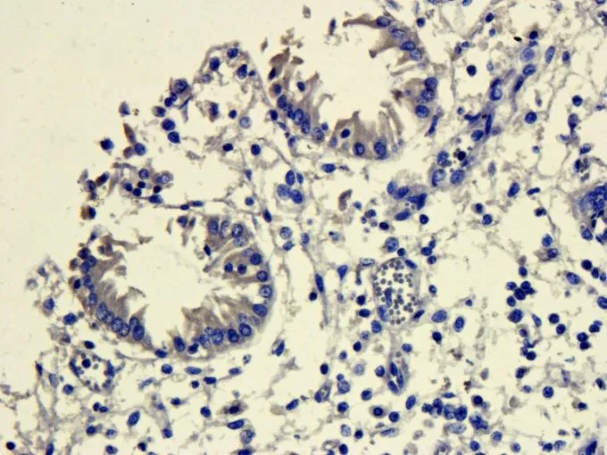
Mannose Receptor antibody [15-2] (Biotin)
GTX28919
ApplicationsFlow Cytometry, ImmunoFluorescence, Western Blot, ImmunoCytoChemistry, ImmunoHistoChemistry, ImmunoHistoChemistry Frozen, ImmunoHistoChemistry Paraffin, Other Application
Product group Antibodies
ReactivityHuman, Mouse, Rat
TargetMRC1
Overview
- SupplierGeneTex
- Product NameMannose Receptor antibody [15-2] (Biotin)
- Delivery Days Customer9
- Application Supplier NoteFor FACS, IA, IHC-Fr and WB: Use at an assay dependent dilution. Optimal dilutions/concentrations should be determined by the researcher.
- ApplicationsFlow Cytometry, ImmunoFluorescence, Western Blot, ImmunoCytoChemistry, ImmunoHistoChemistry, ImmunoHistoChemistry Frozen, ImmunoHistoChemistry Paraffin, Other Application
- CertificationResearch Use Only
- ClonalityMonoclonal
- Clone ID15-2
- Concentration0.1 mg/ml
- ConjugateBiotin
- Gene ID4360
- Target nameMRC1
- Target descriptionmannose receptor C-type 1
- Target synonymsCD206, CLEC13D, CLEC13DL, MMR, MRC1L1, bA541I19.1, hMR, macrophage mannose receptor 1, C-type lectin domain family 13 member D, human mannose receptor, macrophage mannose receptor 1-like protein 1, mannose receptor, C type 1-like 1
- HostMouse
- IsotypeIgG1
- Protein IDP22897
- Protein NameMacrophage mannose receptor 1
- Scientific DescriptionThe recognition of complex carbohydrate structures on glycoproteins is an important part of several biological processes, including cell-cell recognition, serum glycoprotein turnover, and neutralization of pathogens. The protein encoded by this gene is a type I membrane receptor that mediates the endocytosis of glycoproteins by macrophages. The protein has been shown to bind high-mannose structures on the surface of potentially pathogenic viruses, bacteria, and fungi so that they can be neutralized by phagocytic engulfment.[provided by RefSeq, Sep 2015]
- ReactivityHuman, Mouse, Rat
- Storage Instruction2°C to 8°C
- UNSPSC41116161
Datasheet
Related products
Product group Antibodies
MRC1 AntibodyCSB-PA006303
ApplicationsWestern Blot, ELISA
ReactivityHuman
TargetMRC1
- SizePrice
Product group Antibodies
Anti-MRC1 AntibodyA101372
ApplicationsWestern Blot, ELISA
ReactivityHuman
- SizePrice


Product group Antibodies
Anti-MRC1 AntibodyAMAB90746
ApplicationsWestern Blot, ImmunoHistoChemistry
ReactivityHuman
TargetMRC1
- SizePrice
Product group Antibodies

Anti-CD206 [6D5]Ab03382-1.1
ApplicationsELISA
ReactivityHuman
TargetMRC1
- SizePrice

Product group Antibodies
Anti-Mannose Receptor/MRC1 Picoband(r) AntibodyA02285-2-CARRIER-FREE
ApplicationsImmunoFluorescence, Western Blot, ELISA, ImmunoHistoChemistry
ReactivityHuman, Monkey, Mouse, Rat
TargetMRC1
- SizePrice
Product group Antibodies

ApplicationsFlow Cytometry, Western Blot, ImmunoHistoChemistry, ImmunoHistoChemistry Frozen
ReactivityHuman
TargetMRC1
- SizePrice
Product group Antibodies
ApplicationsImmunoPrecipitation, Western Blot, ImmunoCytoChemistry, ImmunoHistoChemistry
ReactivityMouse
TargetMRC1
- SizePrice
Product group Antibodies
References
MRC1 Polyclonal AntibodyBS-4727R
ApplicationsFlow Cytometry, Western Blot, ELISA
ReactivityHuman
TargetMRC1
- SizePrice
Product group Antibodies
References
Mannose Receptor antibodyGTX53806
ApplicationsImmunoHistoChemistry, ImmunoHistoChemistry Paraffin
ReactivityHuman, Mouse, Porcine, Rat
TargetMRC1
- SizePrice