Bio-Connect

Products

Are you looking for life science and diagnostic reagents? We offer one of the most extensive ranges in the Benelux. There are currently more than 5 million products in our webshop, which are manufactured by more than 130 suppliers. This includes both life science and diagnostic reagents. We hope to support your research with everything you need.

1309 to 1320 of 6.750 products
Sort by
Product group Antibodies
Boster Bio
ApplicationsFlow Cytometry, Western Blot, ELISA, ImmunoHistoChemistry
ReactivityHuman, Mouse, Rat
TargetCOL3A1
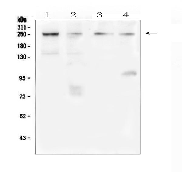
Western blot analysis of TRPM7 using anti-TRPM7 antibody (A00789-1). Electrophoresis was performed on a 5-20% SDS-PAGE gel at 70V (Stacking gel) / 90V (Resolving gel) for 2-3 hours. The sample well of each lane was loaded with 50ug of sample under reducing conditions. Lane 1: human Hela whole cell lysate, Lane 2: human COLO-320 whole cell lysate, Lane 3: human 22RV1 whole cell lysate, Lane 4: human SGC-7901 whole cell lysate. After Electrophoresis, proteins were transferred to a Nitrocellulose membrane at 150mA for 50-90 minutes. Blocked the membrane with 5% Non-fat Milk/ TBS for 1.5 hour at RT. The membrane was incubated with rabbit anti-TRPM7 antigen affinity purified polyclonal antibody (Catalog # A00789-1) at 0.5 μg/mL overnight at 4°C, then washed with TBS-0.1%Tween 3 times with 5 minutes each and probed with a goat anti-rabbit IgG-HRP secondary antibody at a dilution of 1:10000 for 1.5 hour at RT. The signal is developed using an Enhanced Chemiluminescent detection (ECL) kit (Catalog # EK1002) with Tanon 5200 system. A specific band was detected for TRPM7 at approximately 250KD. The expected band size for TRPM7 is at 212KD.
Product group Antibodies
Boster Bio
ApplicationsFlow Cytometry, ImmunoFluorescence, Western Blot, ELISA, ImmunoCytoChemistry
ReactivityHuman, Mouse, Rat
TargetTRPM7
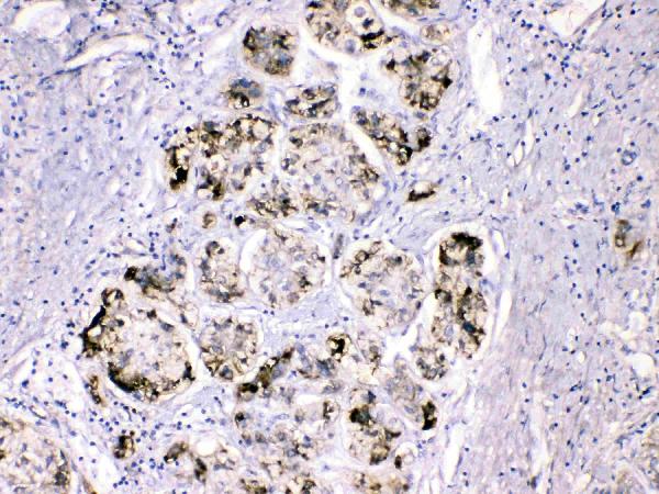
IHC analysis of FGG using anti-FGG antibody (A00790-1).
Product group Antibodies
Boster Bio
ApplicationsFlow Cytometry, Western Blot, ImmunoCytoChemistry, ImmunoHistoChemistry, ImmunoHistoChemistry Frozen
ReactivityHuman, Mouse, Rat
TargetFGG
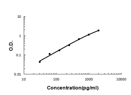
Sandwich ELISA - Recombinant human FGF21 protein standard curve.
Product group Antibodies
Boster Bio
ApplicationsELISA
ReactivityHuman
TargetFGF21
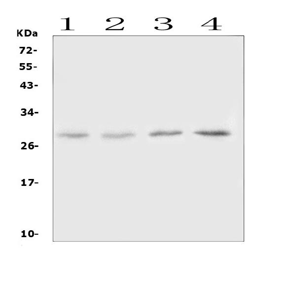
Western blot analysis of Oncostatin M using anti-Oncostatin M antibody (A00804-1). Electrophoresis was performed on a 5-20% SDS-PAGE gel at 70V (Stacking gel) / 90V (Resolving gel) for 2-3 hours. The sample well of each lane was loaded with 50ug of sample under reducing conditions. Lane 1: rat lung tissue lysates, Lane 2: rat spleen tissue lysates, Lane 3: mouse lung tissue lysates, Lane 4: mouse spleen tissue lysates. After Electrophoresis, proteins were transferred to a Nitrocellulose membrane at 150mA for 50-90 minutes. Blocked the membrane with 5% Non-fat Milk/ TBS for 1.5 hour at RT. The membrane was incubated with rabbit anti-Oncostatin M antigen affinity purified polyclonal antibody (Catalog # A00804-1) at 0.5 μg/mL overnight at 4°C, then washed with TBS-0.1%Tween 3 times with 5 minutes each and probed with a goat anti-rabbit IgG-HRP secondary antibody at a dilution of 1:10000 for 1.5 hour at RT. The signal is developed using an Enhanced Chemiluminescent detection (ECL) kit (Catalog # EK1002) with Tanon 5200 system. A specific band was detected for Oncostatin M at approximately 28KD. The expected band size for Oncostatin M is at 28KD.
Product group Antibodies
Boster Bio
ApplicationsWestern Blot, ELISA
ReactivityMouse, Rat
TargetOsm
Western blot analysis of Oncostatin M using anti-Oncostatin M antibody (A00804). Electrophoresis was performed on a 5-20% SDS-PAGE gel at 70V (Stacking gel) / 90V (Resolving gel) for 2-3 hours. The sample well of each lane was loaded with 50ug of sample under reducing conditions. Lane 1: mouse HEPA1-6 whole cell lysates, Lane 2: rat kidney tissue lysates. After Electrophoresis, proteins were transferred to a Nitrocellulose membrane at 150mA for 50-90 minutes. Blocked the membrane with 5% Non-fat Milk/ TBS for 1.5 hour at RT. The membrane was incubated with rabbit anti-Oncostatin M antigen affinity purified polyclonal antibody (Catalog # A00804) at 0.5 ug/mL overnight at 4 then washed with TBS-0.1%Tween 3 times with 5 minutes each and probed with a goat anti-rabbit IgG-HRP secondary antibody at a dilution of 1:10000 for 1.5 hour at RT. The signal is developed using an Enhanced Chemiluminescent detection (ECL) kit (Catalog # EK1002) with Tanon 5200 system. A specific band was detected for Oncostatin M at approximately 28KD. The expected band size for Oncostatin M is at 28KD.
Product group Antibodies
Boster Bio
ApplicationsWestern Blot, ELISA
ReactivityMouse, Rat
TargetOsm
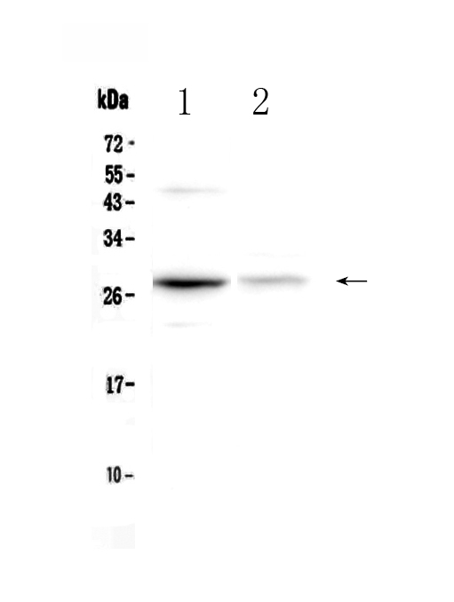
Western blot analysis of LBP using anti-LBP antibody (A00809-1).
Product group Antibodies
Boster Bio
ApplicationsWestern Blot
ReactivityHuman, Mouse, Rat
TargetLBP
Western blot analysis of LBP using anti-LBP antibody (A00809-2). Electrophoresis was performed on a 5-20% SDS-PAGE gel at 70V (Stacking gel) / 90V (Resolving gel) for 2-3 hours. Lane 1: recombinant human LBP protein 1ng. After Electrophoresis, proteins were transferred to a Nitrocellulose membrane at 150mA for 50-90 minutes. Blocked the membrane with 5% Non-fat Milk/ TBS for 1.5 hour at RT. The membrane was incubated with rabbit anti-LBP antigen affinity purified polyclonal antibody (Catalog # A00809-2) at 0.5 ug/mL overnight at 4 then washed with TBS-0.1%Tween 3 times with 5 minutes each and probed with a goat anti-rabbit IgG-HRP secondary antibody at a dilution of 1:10000 for 1.5 hour at RT. The signal is developed using an Enhanced Chemiluminescent detection (ECL) kit (Catalog # EK1002) with Tanon 5200 system. A specific band was detected for LBP at approximately 65KD. The expected band size for LBP is at 51KD.
Product group Antibodies
Boster Bio
ApplicationsWestern Blot, ELISA
ReactivityHuman
TargetLBP
Western blot analysis of LBP using anti-LBP antibody (A00809-3). Electrophoresis was performed on a 5-20% SDS-PAGE gel at 70V (Stacking gel) / 90V (Resolving gel) for 2-3 hours. The sample well of each lane was loaded with 50ug of sample under reducing conditions. Lane 1: human Hela whole cell lysates, Lane 2: human Jurkat whole cell lysates, Lane 3: human A549 whole cell lysates, Lane 4: human Raji whole cell lysates, Lane 5: rat liver tissue lysates, Lane 6: mouse liver tissue lysates. After Electrophoresis, proteins were transferred to a Nitrocellulose membrane at 150mA for 50-90 minutes. Blocked the membrane with 5% Non-fat Milk/ TBS for 1.5 hour at RT. The membrane was incubated with rabbit anti-LBP antigen affinity purified polyclonal antibody (Catalog # A00809-3) at 0.5 μg/mL overnight at 4°C, then washed with TBS-0.1%Tween 3 times with 5 minutes each and probed with a goat anti-rabbit IgG-HRP secondary antibody at a dilution of 1:10000 for 1.5 hour at RT. The signal is developed using an Enhanced Chemiluminescent detection (ECL) kit (Catalog # EK1002) with Tanon 5200 system. A specific band was detected for LBP at approximately 65KD. The expected band size for LBP is at 53KD.
Product group Antibodies
Boster Bio
ApplicationsWestern Blot, ELISA
ReactivityHuman, Mouse, Rat
TargetLbp
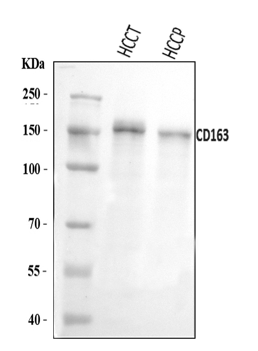
Western blot analysis of CD163 using anti-CD163 antibody (A00812-1).
Product group Antibodies
References
Boster Bio
ApplicationsWestern Blot, ELISA, ImmunoHistoChemistry
ReactivityHuman
TargetCD163
Boster Kit Box
Product group Antibodies
Boster Bio
ApplicationsFlow Cytometry
ReactivityHuman
TargetCD163
Boster Kit Box
Product group Antibodies
Boster Bio
ApplicationsFlow Cytometry
ReactivityHuman
TargetCD163