

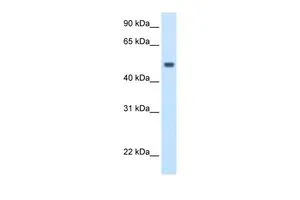
Various tissue extracts (50 μg) were separated by 10% SDS-PAGE, and the membrane was blotted with MAT1A + MAT2A antibody [HL1679] (GTX637278) diluted at 1:1000. The HRP-conjugated anti-rabbit IgG antibody (GTX213110-01) was used to detect the primary antibody.
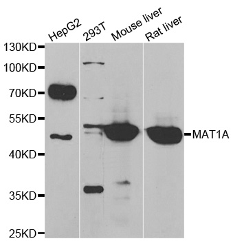
![Various whole cell extracts (30 μg) were separated by 10% SDS-PAGE, and the membrane was blotted with MAT1A + MAT2A antibody [HL1679] (GTX637278) diluted at 1:2000. The HRP-conjugated anti-rabbit IgG antibody (GTX213110-01) was used to detect the primary antibody.](https://www.genetex.com/upload/website/prouct_img/normal/GTX637278/GTX637278_44865_20221118_WB_22112219_177.webp "Various whole cell extracts (30 μg) were separated by 10% SDS-PAGE, and the membrane was blotted with MAT1A + MAT2A antibody [HL1679] (GTX637278) diluted at 1:2000. The HRP-conjugated anti-rabbit IgG antibody (GTX213110-01) was used to detect the primary antibody.")
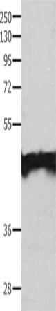
![293T whole cell extracts (30 μg) were separated by 10% SDS-PAGE, and the membrane was blotted with MAT1A + MAT2A antibody [HL1679] (GTX637278) diluted at 1:5000. The HRP-conjugated anti-rabbit IgG antibody (GTX213110-01) was used to detect the primary antibody.](https://www.genetex.com/upload/website/prouct_img/normal/GTX637278/GTX637278_44865_20221125_WB_B_22112723_766.webp "293T whole cell extracts (30 μg) were separated by 10% SDS-PAGE, and the membrane was blotted with MAT1A + MAT2A antibody [HL1679] (GTX637278) diluted at 1:5000. The HRP-conjugated anti-rabbit IgG antibody (GTX213110-01) was used to detect the primary antibody.")
![Drosophila tissue extract (50 μg) was separated by 10% SDS-PAGE, and the membrane was blotted with MAT1A + MAT2A antibody [HL1679] (GTX637278) diluted at 1:1000. The HRP-conjugated anti-rabbit IgG antibody (GTX213110-01) was used to detect the primary antibody.](https://www.genetex.com/upload/website/prouct_img/normal/GTX637278/GTX637278_44865_20230324_WB_Drosophila_brain_23032819_179.webp "Drosophila tissue extract (50 μg) was separated by 10% SDS-PAGE, and the membrane was blotted with MAT1A + MAT2A antibody [HL1679] (GTX637278) diluted at 1:1000. The HRP-conjugated anti-rabbit IgG antibody (GTX213110-01) was used to detect the primary antibody.")
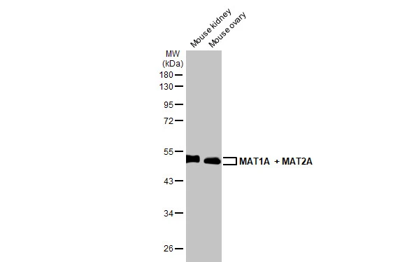
Various tissue extracts (50 μg) were separated by 10% SDS-PAGE, and the membrane was blotted with MAT1A + MAT2A antibody [HL1679] (GTX637278) diluted at 1:1000. The HRP-conjugated anti-rabbit IgG antibody (GTX213110-01) was used to detect the primary antibody.
MAT1A + MAT2A antibody [HL1679]
GTX637278
ApplicationsWestern Blot
Product group Antibodies
ReactivityDrosophila, Human, Mouse
TargetMAT1A
Overview
- SupplierGeneTex
- Product NameMAT1A + MAT2A antibody [HL1679]
- Delivery Days Customer9
- Application Supplier NoteWB: 1:500-1:3000. *Optimal dilutions/concentrations should be determined by the researcher.Not tested in other applications.
- ApplicationsWestern Blot
- CertificationResearch Use Only
- ClonalityMonoclonal
- Clone IDHL1679
- Concentration1 mg/ml
- ConjugateUnconjugated
- Gene ID4143
- Target nameMAT1A
- Target descriptionmethionine adenosyltransferase 1A
- Target synonymsMAT, MATA1, SAMS, SAMS1, S-adenosylmethionine synthase isoform type-1, MAT 1, MAT-I/III, adoMet synthase 1, adoMet synthetase 1, methionine adenosyltransferase 1, methionine adenosyltransferase I, alpha, methionine adenosyltransferase I/III
- HostRabbit
- IsotypeIgG
- Protein IDP31153
- Protein NameS-adenosylmethionine synthase isoform type-2
- ReactivityDrosophila, Human, Mouse
- Storage Instruction-20°C or -80°C,2°C to 8°C
- UNSPSC41116161
Datasheet
Related products
Product group Antibodies
Anti-MAT1A AntibodyA31491
ApplicationsWestern Blot, ImmunoHistoChemistry
ReactivityHuman, Mouse, Rat
- SizePrice

Product group Antibodies
Anti-MAT1A Antibody Picoband(r)A04203-1-CARRIER-FREE
ApplicationsWestern Blot
ReactivityMouse, Rat
TargetMAT1A
- SizePrice
Product group Antibodies
Anti-MAT1A Antibody144-06650
ApplicationsWestern Blot
ReactivityHuman, Mouse, Rat
TargetMAT1A
- SizePrice
Product group Antibodies

MAT / MAT1A AntibodyLS-C746819
ApplicationsWestern Blot, ImmunoHistoChemistry
ReactivityHuman, Mouse, Rat
TargetMAT1A
- SizePrice
Product group Antibodies
References
MAT2A Polyclonal AntibodyBS-4054R
ApplicationsImmunoFluorescence, Western Blot, ELISA, ImmunoCytoChemistry, ImmunoHistoChemistry, ImmunoHistoChemistry Frozen, ImmunoHistoChemistry Paraffin
ReactivityBovine, Canine, Chicken, Human, Monkey, Mouse, Porcine, Rabbit, Rat, Zebra Fish
TargetMAT1A
- SizePrice
Product group Antibodies
MAT1A AntibodyCSB-PA231283
ApplicationsWestern Blot, ELISA
ReactivityHuman, Mouse, Rat
TargetMAT1A
- SizePrice
Product group Antibodies
MAT1A + MAT2A antibodyGTX132095
ApplicationsWestern Blot, ImmunoHistoChemistry, ImmunoHistoChemistry Paraffin
ReactivityHuman, Mouse, Rat
TargetMAT1A
- SizePrice

Product group Antibodies
Anti-MAT1A AntibodyHPA048627
ApplicationsImmunoHistoChemistry
ReactivityHuman
TargetMAT1A
- SizePrice
Product group Antibodies
MAT1A antibody, C-termGTX47162
ApplicationsWestern Blot
ReactivityHuman
TargetMAT1A
- SizePrice
Product group Antibodies
References
MAT1A antibody, N-termGTX47163
ApplicationsWestern Blot, ImmunoHistoChemistry, ImmunoHistoChemistry Paraffin
ReactivityHuman
TargetMAT1A
- SizePrice