

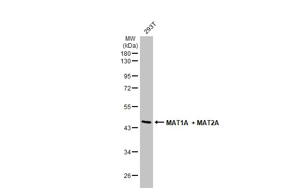
Various whole cell extracts (30 μg) was separated by 10% SDS-PAGE, and the membrane was blotted with MAT1A + MAT2A antibody (GTX112535) diluted at 1:2000. The HRP-conjugated anti-rabbit IgG antibody (GTX213110-01) was used to detect the primary antibody.
 were separated by 10% SDS-PAGE, and the membrane was blotted with MAT1A + MAT2A antibody (GTX112535) diluted at 1:2000. The HRP-conjugated anti-rabbit IgG antibody (GTX213110-01) was used to detect the primary antibody.Corresponding RNA expression data for the same cell lines are based on Human Protein Atlas program.")
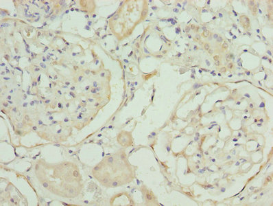
![MAT1A + MAT2A antibody [N1C2] detects MAT1A + MAT2A protein at cytoplasm and nucleus by immunohistochemical analysis. Sample: Paraffin-embedded rat colon. MAT1A + MAT2A stained by MAT1A + MAT2A antibody [N1C2] (GTX112535) diluted at 1:500. Antigen Retrieval: Citrate buffer, pH 6.0, 15 min](https://www.genetex.com/upload/website/prouct_img/normal/GTX112535/GTX112535_44609_20220506_IHC-P_R_1_w_23060500_144.webp "MAT1A + MAT2A antibody [N1C2] detects MAT1A + MAT2A protein at cytoplasm and nucleus by immunohistochemical analysis. Sample: Paraffin-embedded rat colon. MAT1A + MAT2A stained by MAT1A + MAT2A antibody [N1C2] (GTX112535) diluted at 1:500. Antigen Retrieval: Citrate buffer, pH 6.0, 15 min")
![MAT1A + MAT2A antibody [N1C2] detects MAT1A + MAT2A protein at endoplasmic reticulum by immunofluorescent analysis. Sample: HeLa cells were fixed in 4% paraformaldehyde at RT for 15 min. Green: MAT1A + MAT2A stained by MAT1A + MAT2A antibody [N1C2] (GTX112535) diluted at 1:500. Blue: Fluoroshield with DAPI (GTX30920).](https://www.genetex.com/upload/website/prouct_img/normal/GTX112535/GTX112535_44609_20220429_ICC_IF_w_23060500_991.webp "MAT1A + MAT2A antibody [N1C2] detects MAT1A + MAT2A protein at endoplasmic reticulum by immunofluorescent analysis. Sample: HeLa cells were fixed in 4% paraformaldehyde at RT for 15 min. Green: MAT1A + MAT2A stained by MAT1A + MAT2A antibody [N1C2] (GTX112535) diluted at 1:500. Blue: Fluoroshield with DAPI (GTX30920).")
![MAT1A + MAT2A antibody [N1C2] detects MAT1A + MAT2A protein at cytoplasm and nucleus by immunohistochemical analysis. Sample: Paraffin-embedded rat colon. MAT1A + MAT2A stained by MAT1A + MAT2A antibody [N1C2] (GTX112535) diluted at 1:500. Antigen Retrieval: Citrate buffer, pH 6.0, 15 min](https://www.genetex.com/upload/website/prouct_img/normal/GTX112535/GTX112535_44609_20220506_IHC-P_R_w_23060500_936.webp "MAT1A + MAT2A antibody [N1C2] detects MAT1A + MAT2A protein at cytoplasm and nucleus by immunohistochemical analysis. Sample: Paraffin-embedded rat colon. MAT1A + MAT2A stained by MAT1A + MAT2A antibody [N1C2] (GTX112535) diluted at 1:500. Antigen Retrieval: Citrate buffer, pH 6.0, 15 min")
![MAT1A + MAT2A antibody [N1C2] detects MAT1A + MAT2A protein at cytoplasm and nucleus by immunohistochemical analysis. Sample: Paraffin-embedded mouse esophagus. MAT1A + MAT2A stained by MAT1A + MAT2A antibody [N1C2] (GTX112535) diluted at 1:500. Antigen Retrieval: Citrate buffer, pH 6.0, 15 min](https://www.genetex.com/upload/website/prouct_img/normal/GTX112535/GTX112535_44609_20220506_IHC-P_M_w_23060500_691.webp "MAT1A + MAT2A antibody [N1C2] detects MAT1A + MAT2A protein at cytoplasm and nucleus by immunohistochemical analysis. Sample: Paraffin-embedded mouse esophagus. MAT1A + MAT2A stained by MAT1A + MAT2A antibody [N1C2] (GTX112535) diluted at 1:500. Antigen Retrieval: Citrate buffer, pH 6.0, 15 min")
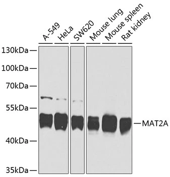
Various whole cell extracts (30 μg) was separated by 10% SDS-PAGE, and the membrane was blotted with MAT1A + MAT2A antibody (GTX112535) diluted at 1:2000. The HRP-conjugated anti-rabbit IgG antibody (GTX213110-01) was used to detect the primary antibody.
MAT1A + MAT2A antibody [N1C2]
GTX112535
ApplicationsImmunoFluorescence, Western Blot, ImmunoCytoChemistry, ImmunoHistoChemistry, ImmunoHistoChemistry Paraffin
Product group Antibodies
ReactivityHuman, Mouse, Rat
TargetMAT2A
Overview
- SupplierGeneTex
- Product NameMAT1A + MAT2A antibody [N1C2]
- Delivery Days Customer9
- Application Supplier NoteWB: 1:500-1:3000. *Optimal dilutions/concentrations should be determined by the researcher.Not tested in other applications.
- ApplicationsImmunoFluorescence, Western Blot, ImmunoCytoChemistry, ImmunoHistoChemistry, ImmunoHistoChemistry Paraffin
- CertificationResearch Use Only
- ClonalityPolyclonal
- Concentration0.18 mg/ml
- ConjugateUnconjugated
- Gene ID4144
- Target nameMAT2A
- Target descriptionmethionine adenosyltransferase 2A
- Target synonymsMATA2, MATII, SAMS2, S-adenosylmethionine synthase isoform type-2, MAT 2, MAT-II, adoMet synthase 2, adoMet synthetase 2, methionine adenosyltransferase 2, methionine adenosyltransferase II, alpha, testicular tissue protein Li 121
- HostRabbit
- IsotypeIgG
- Protein IDP31153
- Protein NameS-adenosylmethionine synthase isoform type-2
- Scientific DescriptionCatalyzes the formation of S-adenosylmethionine from methionine and ATP.
- ReactivityHuman, Mouse, Rat
- Storage Instruction-20°C or -80°C,2°C to 8°C
- UNSPSC41116161
Datasheet
Related products
Product group Antibodies
MAT2A AntibodyCSB-PA013517LA01HU
ApplicationsImmunoFluorescence, ELISA, ImmunoHistoChemistry
ReactivityHuman
TargetMAT2A
- SizePrice

Product group Antibodies
Anti-MAT2A AntibodyA16194
ApplicationsWestern Blot
ReactivityHuman, Mouse, Rat
- SizePrice

Product group Antibodies
Anti-MAT2A-25ulHPA043028
ApplicationsWestern Blot, ImmunoCytoChemistry, ImmunoHistoChemistry
ReactivityHuman
- SizePrice

Product group Antibodies
Anti-MAT2A Antibody Picoband(r)A04557-1-CARRIER-FREE
ApplicationsFlow Cytometry, Western Blot, ELISA, ImmunoHistoChemistry
ReactivityHuman, Mouse, Rat
TargetMAT2A
- SizePrice
Product group Antibodies

MAT2A AntibodyLS-C409971
ApplicationsWestern Blot
ReactivityHuman, Mouse, Rat
TargetMAT2A
- SizePrice
Product group Antibodies
ApplicationsImmunoFluorescence, Western Blot, ImmunoCytoChemistry, ImmunoHistoChemistry
TargetMAT2A
- SizePrice
Product group Antibodies
MAT2A antibodyGTX64527
ApplicationsWestern Blot
ReactivityHuman, Mouse, Rat
TargetMAT2A
- SizePrice
Product group Antibodies
ApplicationsWestern Blot
ReactivityBovine, Human, Primate, Rat, Zebra Fish
TargetMAT2A
- SizePrice
Product group Antibodies
ApplicationsWestern Blot
ReactivityBovine, Human, Primate, Rat, Zebra Fish
TargetMAT2A
- SizePrice