

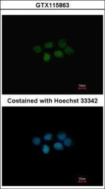
Immunofluorescence analysis of paraformaldehyde-fixed A431, using MAT2B(GTX115863) antibody at 1:200 dilution.
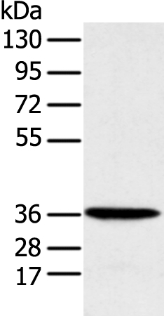
![Wild-type (WT) and MAT2B knockout (KO) 293T cell extracts (30 μg) were separated by 10% SDS-PAGE, and the membrane was blotted with MAT2B antibody [N3C3] (GTX115863) diluted at 1:1000. The HRP-conjugated anti-rabbit IgG antibody (GTX213110-01) was used to detect the primary antibody, and the signal was developed with Trident ECL plus-Enhanced.](https://www.genetex.com/upload/website/prouct_img/normal/GTX115863/GTX115863_40296_20180316_WB_KO_watermark_w_23060519_542.webp "Wild-type (WT) and MAT2B knockout (KO) 293T cell extracts (30 μg) were separated by 10% SDS-PAGE, and the membrane was blotted with MAT2B antibody [N3C3] (GTX115863) diluted at 1:1000. The HRP-conjugated anti-rabbit IgG antibody (GTX213110-01) was used to detect the primary antibody, and the signal was developed with Trident ECL plus-Enhanced.")
Immunofluorescence analysis of paraformaldehyde-fixed A431, using MAT2B(GTX115863) antibody at 1:200 dilution.
MAT2B antibody [N3C3]
GTX115863
ApplicationsImmunoFluorescence, Western Blot, ImmunoCytoChemistry
Product group Antibodies
ReactivityHuman, Mouse
TargetMAT2B
Overview
- SupplierGeneTex
- Product NameMAT2B antibody [N3C3]
- Delivery Days Customer9
- Application Supplier NoteWB: 1:500-1:3000. ICC/IF: 1:100-1:1000. *Optimal dilutions/concentrations should be determined by the researcher.Not tested in other applications.
- ApplicationsImmunoFluorescence, Western Blot, ImmunoCytoChemistry
- CertificationResearch Use Only
- ClonalityPolyclonal
- Concentration0.45 mg/ml
- ConjugateUnconjugated
- Gene ID27430
- Target nameMAT2B
- Target descriptionmethionine adenosyltransferase 2 non-catalytic beta subunit
- Target synonymsMAT-II, MATIIbeta, Nbla02999, SDR23E1, TGR, methionine adenosyltransferase 2 subunit beta, MAT II beta, beta regulatory subunit of methionine adenosyltransferase, dTDP-4-keto-6-deoxy-D-glucose 4-reductase, methionine adenosyltransferase 2B, methionine adenosyltransferase II, beta, putative dTDP-4-keto-6-deoxy-D-glucose 4-reductase, putative protein product of Nbla02999, short chain dehydrogenase/reductase family 23E, member 1, testicular tissue protein Li 118
- HostRabbit
- IsotypeIgG
- Protein IDQ9NZL9
- Protein NameMethionine adenosyltransferase 2 subunit beta
- Scientific DescriptionThe protein encoded by this gene belongs to the methionine adenosyltransferase (MAT) family. MAT catalyzes the biosynthesis of S-adenosylmethionine from methionine and ATP. This protein is the regulatory beta subunit of MAT. Alternative splicing occurs at this locus and two transcript variants encoding distinct isoforms have been identified. [provided by RefSeq]
- ReactivityHuman, Mouse
- Storage Instruction-20°C or -80°C,2°C to 8°C
- UNSPSC12352203
References
- Fisslthaler B, Zippel N, Abdel Malik R, et al. Myeloid-Specific Deletion of the AMPKα2 Subunit Alters Monocyte Protein Expression and Atherogenesis. Int J Mol Sci. 2019,20(12). doi: 10.3390/ijms20123005Read this paper
Datasheet
Related products
Product group Antibodies
Anti-MAT2B Antibody144-11608
ApplicationsWestern Blot
ReactivityHuman, Mouse
TargetMAT2B
- SizePrice
Product group Antibodies
MAT2B Polyclonal AntibodyCAC14799
ApplicationsImmunoFluorescence, Western Blot, ELISA
ReactivityRat
TargetMAT2B
- SizePrice
Product group Antibodies
MAT2B antibodyGTX64937
ApplicationsWestern Blot
ReactivityHuman, Mouse
TargetMAT2B
- SizePrice
Product group Antibodies

Goat anti-MAT2B (isoform 1)EB11612
ApplicationsWestern Blot, ELISA
ReactivityBovine, Canine, Human, Mouse, Porcine, Rat
TargetMAT2B
- SizePrice

Product group Antibodies
Anti-MAT2B/TGR Antibody Picoband(r)A06719-2-CARRIER-FREE
ApplicationsFlow Cytometry, Western Blot, ELISA
ReactivityHuman
TargetMAT2B
- SizePrice
Product group Antibodies

MAT2B AntibodyLS-C830335
ApplicationsWestern Blot, ELISA
ReactivityHuman, Mouse, Rat
TargetMAT2B
- SizePrice
Product group Antibodies
MAT2B AntibodyCSB-PA882157LA01HU
ApplicationsImmunoFluorescence, Western Blot, ELISA
ReactivityHuman, Rat
TargetMAT2B
- SizePrice

Product group Antibodies
Anti-MAT2B AntibodyHPA037722
ApplicationsWestern Blot, ImmunoHistoChemistry
ReactivityHuman
TargetMAT2B
- SizePrice