

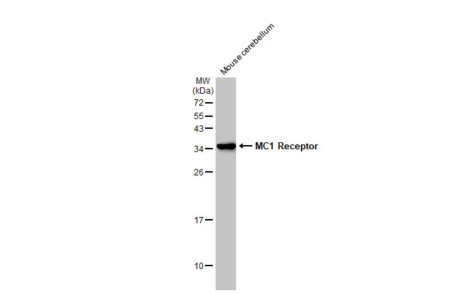
Mouse tissue extract (50 μg) was separated by 12% SDS-PAGE, and the membrane was blotted with MC1 Receptor antibody [C2C3], C-term (GTX108190) diluted at 1:4000. The HRP-conjugated anti-rabbit IgG antibody (GTX213110-01) was used to detect the primary antibody.
![MC1 Receptor antibody [C2C3], C-term detects MC1R protein at membrane on mouse fore brain by immunohistochemical analysis. Sample: Paraffin-embedded mouse fore brain. MC1 Receptor antibody [C2C3], C-term (GTX108190) dilution: 1:500.
Antigen Retrieval: Trilogy? (EDTA based, pH 8.0) buffer, 15min](https://www.genetex.com/upload/website/prouct_img/normal/GTX108190/GTX108190_39764_IHC_M_w_23060120_337.webp "MC1 Receptor antibody [C2C3], C-term detects MC1R protein at membrane on mouse fore brain by immunohistochemical analysis. Sample: Paraffin-embedded mouse fore brain. MC1 Receptor antibody [C2C3], C-term (GTX108190) dilution: 1:500.
Antigen Retrieval: Trilogy? (EDTA based, pH 8.0) buffer, 15min")
![MC1 Receptor antibody [C2C3], C-term detects MC1R protein at cytosol and membrane on human colon carcinoma by immunohistochemical analysis. Sample: Paraffin-embedded colon carcinoma. MC1 Receptor antibody [C2C3], C-term (GTX108190) dilution: 1:500.
Antigen Retrieval: Trilogy? (EDTA based, pH 8.0) buffer, 15min](https://www.genetex.com/upload/website/prouct_img/normal/GTX108190/GTX108190_39764_IHC_w_23060120_644.webp "MC1 Receptor antibody [C2C3], C-term detects MC1R protein at cytosol and membrane on human colon carcinoma by immunohistochemical analysis. Sample: Paraffin-embedded colon carcinoma. MC1 Receptor antibody [C2C3], C-term (GTX108190) dilution: 1:500.
Antigen Retrieval: Trilogy? (EDTA based, pH 8.0) buffer, 15min")
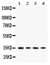
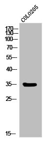
![Whole cell extract (30 μg) was separated by 12% SDS-PAGE, and the membrane was blotted with MC1 Receptor antibody [C2C3], C-term (GTX108190) diluted at 1:4000. The HRP-conjugated anti-rabbit IgG antibody (GTX213110-01) was used to detect the primary antibody.](https://www.genetex.com/upload/website/prouct_img/normal/GTX108190/GTX108190_44916_20230120_WB_24070822_790.webp "Whole cell extract (30 μg) was separated by 12% SDS-PAGE, and the membrane was blotted with MC1 Receptor antibody [C2C3], C-term (GTX108190) diluted at 1:4000. The HRP-conjugated anti-rabbit IgG antibody (GTX213110-01) was used to detect the primary antibody.")
Mouse tissue extract (50 μg) was separated by 12% SDS-PAGE, and the membrane was blotted with MC1 Receptor antibody [C2C3], C-term (GTX108190) diluted at 1:4000. The HRP-conjugated anti-rabbit IgG antibody (GTX213110-01) was used to detect the primary antibody.
MC1 Receptor antibody [C2C3], C-term
GTX108190
ApplicationsImmunoFluorescence, Western Blot, ImmunoCytoChemistry, ImmunoHistoChemistry, ImmunoHistoChemistry Frozen, ImmunoHistoChemistry Paraffin
Product group Antibodies
ReactivityHuman, Mouse
TargetMC1R
Overview
- SupplierGeneTex
- Product NameMC1 Receptor antibody [C2C3], C-term
- Delivery Days Customer9
- Application Supplier NoteWB: 1:500-1:3000. IHC-P: 1:100-1:1000. *Optimal dilutions/concentrations should be determined by the researcher.Not tested in other applications.
- ApplicationsImmunoFluorescence, Western Blot, ImmunoCytoChemistry, ImmunoHistoChemistry, ImmunoHistoChemistry Frozen, ImmunoHistoChemistry Paraffin
- CertificationResearch Use Only
- ClonalityPolyclonal
- Concentration0.58 mg/ml
- ConjugateUnconjugated
- Gene ID4157
- Target nameMC1R
- Target descriptionmelanocortin 1 receptor
- Target synonymsCMM5, MSH-R, SHEP2, melanocyte-stimulating hormone receptor, MC1-R, alpha melanocyte stimulating hormone receptor, melanotropin receptor
- HostRabbit
- IsotypeIgG
- Protein IDQ01726
- Protein NameMelanocyte-stimulating hormone receptor
- Scientific DescriptionThis intronless gene encodes the receptor protein for melanocyte-stimulating hormone (MSH). The encoded protein, a seven pass transmembrane G protein coupled receptor, controls melanogenesis. Two types of melanin exist: red pheomelanin and black eumelanin. Gene mutations that lead to a loss in function are associated with increased pheomelanin production, which leads to lighter skin and hair color. Eumelanin is photoprotective but pheomelanin may contribute to UV-induced skin damage by generating free radicals upon UV radiation. Binding of MSH to its receptor activates the receptor and stimulates eumelanin synthesis. This receptor is a major determining factor in sun sensitivity and is a genetic risk factor for melanoma and non-melanoma skin cancer. Over 30 variant alleles have been identified which correlate with skin and hair color, providing evidence that this gene is an important component in determining normal human pigment variation. [provided by RefSeq]
- ReactivityHuman, Mouse
- Storage Instruction-20°C or -80°C,2°C to 8°C
- UNSPSC41116161
Datasheet
Related products
Product group Antibodies
Anti-MC1R AntibodyA38879
ApplicationsWestern Blot, ImmunoHistoChemistry
ReactivityHuman, Mouse
- SizePrice
Product group Antibodies
Anti-MC1 Receptor/MC1R Antibody Picoband(r)A00855-1-CARRIER-FREE
ApplicationsWestern Blot
ReactivityHuman, Mouse, Rat
TargetMC1R
- SizePrice
Product group Antibodies
Anti-MC1R Antibody144-60215
ApplicationsWestern Blot
ReactivityHuman, Mouse, Rat
TargetMC1R
- SizePrice
Product group Antibodies
MC1R Polyclonal AntibodyBS-23517R
ApplicationsImmunoFluorescence, Western Blot, ELISA, ImmunoCytoChemistry, ImmunoHistoChemistry, ImmunoHistoChemistry Frozen, ImmunoHistoChemistry Paraffin
ReactivityHuman
TargetMC1R
- SizePrice
Product group Antibodies
MC1R AntibodyCSB-PA010093
ApplicationsImmunoFluorescence, Western Blot, ELISA
ReactivityHuman
TargetMC1R
- SizePrice
Product group Antibodies
Mc1R Polyclonal AntibodyCAC11237
ApplicationsImmunoFluorescence, Western Blot, ELISA, ImmunoHistoChemistry
TargetMC1R
- SizePrice
Product group Antibodies

MC1R AntibodyLS-C402431
ApplicationsWestern Blot, ELISA
ReactivityHuman, Mouse
TargetMC1R
- SizePrice
![Various whole cell extracts (30 μg) were separated by 12% SDS-PAGE, and the membrane was blotted with MC1 Receptor antibody [HL1470] (GTX636944) diluted at 1:100000. The HRP-conjugated anti-rabbit IgG antibody (GTX213110-01) was used to detect the primary antibody. Corresponding RNA expression data for the same cell lines are based on Human Protein Atlas program.](https://www.genetex.com/upload/website/prouct_img/normal/GTX636944/GTX636944_44711_20230317_WB_TPM_watermark_23032022_421.webp)
Product group Antibodies
MC1 Receptor antibody [HL1470]GTX636944
ApplicationsImmunoFluorescence, Western Blot, ImmunoCytoChemistry
ReactivityHuman, Mouse, Rat
TargetMC1R
- SizePrice
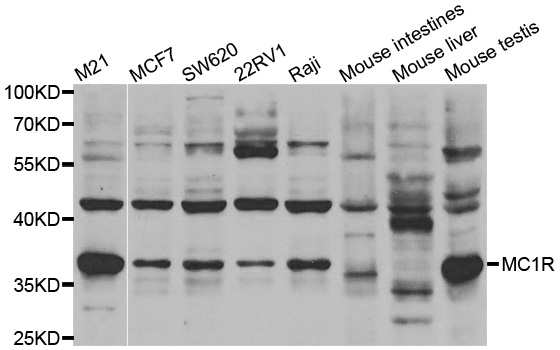
![Various tissue extracts (50 μg) were separated by 12% SDS-PAGE, and the membrane was blotted with MC1 Receptor antibody [HL1471] (GTX636945) diluted at 1:1000. The HRP-conjugated anti-rabbit IgG antibody (GTX213110-01) was used to detect the primary antibody.](https://www.genetex.com/upload/website/prouct_img/normal/GTX636945/GTX636945_T-44662_20230616_WB_M_tissue_23062019_114.webp)
Product group Antibodies
MC1 Receptor antibody [HL1471]GTX636945
ApplicationsImmunoFluorescence, Western Blot, ImmunoCytoChemistry, ImmunoHistoChemistry, ImmunoHistoChemistry Frozen, ImmunoHistoChemistry Paraffin
ReactivityHuman, Mouse, Rat
TargetMC1R
- SizePrice