

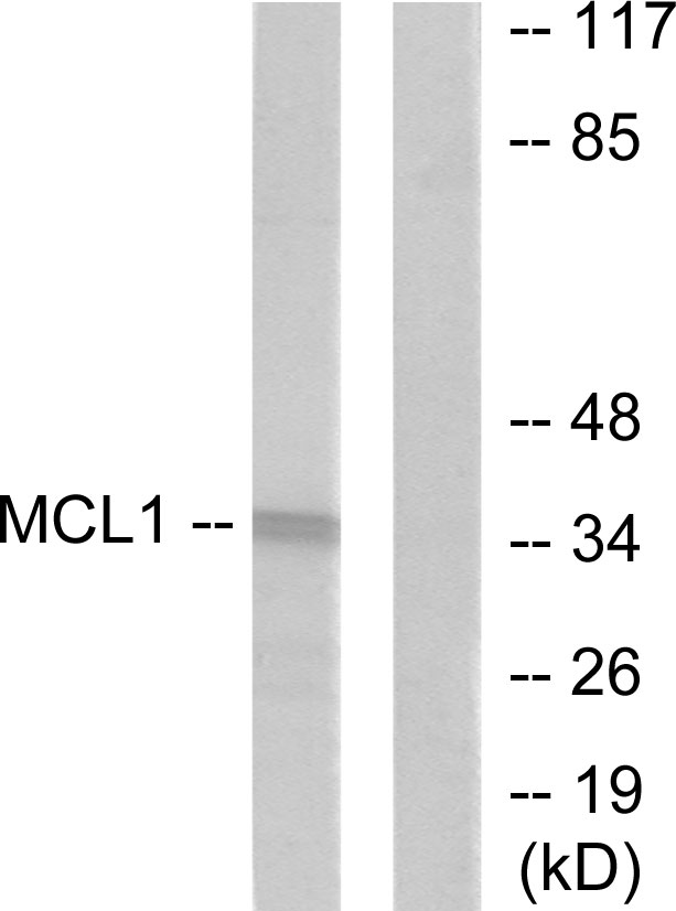
Various whole cell extracts (30 μg) were separated by 10% SDS-PAGE, and the membrane was blotted with MCL1 antibody [HL1544] (GTX637019) diluted at 1:1000. The HRP-conjugated anti-rabbit IgG antibody (GTX213110-01) was used to detect the primary antibody.
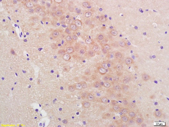
![MCL1 antibody [HL1544] detects MCL1 protein at cytoplasm by immunohistochemical analysis. Sample: Paraffin-embedded human breast carcinoma. MCL1 stained by MCL1 antibody [HL1544] (GTX637019) diluted at 1:100. Antigen Retrieval: Citrate buffer, pH 6.0, 15 min](https://www.genetex.com/upload/website/prouct_img/normal/GTX637019/GTX637019_T-44697_20220624_IHC-P_22062919_337.webp "MCL1 antibody [HL1544] detects MCL1 protein at cytoplasm by immunohistochemical analysis. Sample: Paraffin-embedded human breast carcinoma. MCL1 stained by MCL1 antibody [HL1544] (GTX637019) diluted at 1:100. Antigen Retrieval: Citrate buffer, pH 6.0, 15 min")
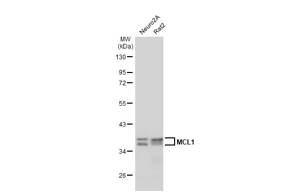
![Various whole cell extracts (30 μg) were separated by 10% SDS-PAGE, and the membrane was blotted with MCL1 antibody [HL1544] (GTX637019) diluted at 1:1000. The HRP-conjugated anti-rabbit IgG antibody (GTX213110-01) was used to detect the primary antibody.](https://www.genetex.com/upload/website/prouct_img/normal/GTX637019/GTX637019_44746_20220722_WB_22072519_335.webp "Various whole cell extracts (30 μg) were separated by 10% SDS-PAGE, and the membrane was blotted with MCL1 antibody [HL1544] (GTX637019) diluted at 1:1000. The HRP-conjugated anti-rabbit IgG antibody (GTX213110-01) was used to detect the primary antibody.")
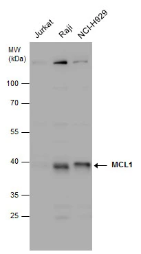
![Various whole cell extracts (30 μg) were separated by 10% SDS-PAGE, and the membrane was blotted with MCL1 antibody [HL1544] (GTX637019) diluted at 1:1000. The HRP-conjugated anti-rabbit IgG antibody (GTX213110-01) was used to detect the primary antibody. Corresponding RNA expression data for the same cell lines are based on Human Protein Atlas program.](https://www.genetex.com/upload/website/prouct_img/normal/GTX637019/GTX637019_44746_20220812_WB_TPM_watermark_22081423_970.webp "Various whole cell extracts (30 μg) were separated by 10% SDS-PAGE, and the membrane was blotted with MCL1 antibody [HL1544] (GTX637019) diluted at 1:1000. The HRP-conjugated anti-rabbit IgG antibody (GTX213110-01) was used to detect the primary antibody. Corresponding RNA expression data for the same cell lines are based on Human Protein Atlas program.")
![MCL1 antibody [HL1544] detects MCL1 protein by immunohistochemical analysis. Sample: Paraffin-embedded mouse tissues. MCL1 stained by MCL1 antibody [HL1544] (GTX637019) diluted at 1:100. Antigen Retrieval: Citrate buffer, pH 6.0, 15 min](https://www.genetex.com/upload/website/prouct_img/normal/GTX637019/GTX637019_44746_20221227_IHC-P_multiple_M_22122821_984.webp "MCL1 antibody [HL1544] detects MCL1 protein by immunohistochemical analysis. Sample: Paraffin-embedded mouse tissues. MCL1 stained by MCL1 antibody [HL1544] (GTX637019) diluted at 1:100. Antigen Retrieval: Citrate buffer, pH 6.0, 15 min")
![MCL1 antibody [HL1544] detects MCL1 protein by immunohistochemical analysis. Sample: Paraffin-embedded dog tissue. MCL1 stained by MCL1 antibody [HL1544] (GTX637019) diluted at 1:100. Antigen Retrieval: Citrate buffer, pH 6.0, 15 min](https://www.genetex.com/upload/website/prouct_img/normal/GTX637019/GTX637019_44746_20230217_IHC-P_multiple_Dog_23030219_481.webp "MCL1 antibody [HL1544] detects MCL1 protein by immunohistochemical analysis. Sample: Paraffin-embedded dog tissue. MCL1 stained by MCL1 antibody [HL1544] (GTX637019) diluted at 1:100. Antigen Retrieval: Citrate buffer, pH 6.0, 15 min")
![MCL1 antibody [HL1544] detects MCL1 protein by immunohistochemical analysis. Sample: Paraffin-embedded cat tissue. MCL1 stained by MCL1 antibody [HL1544] (GTX637019) diluted at 1:100. Antigen Retrieval: Citrate buffer, pH 6.0, 15 min](https://www.genetex.com/upload/website/prouct_img/normal/GTX637019/GTX637019_44746_20230217_IHC-P_multiple_Cat_23030219_596.webp "MCL1 antibody [HL1544] detects MCL1 protein by immunohistochemical analysis. Sample: Paraffin-embedded cat tissue. MCL1 stained by MCL1 antibody [HL1544] (GTX637019) diluted at 1:100. Antigen Retrieval: Citrate buffer, pH 6.0, 15 min")
Various whole cell extracts (30 μg) were separated by 10% SDS-PAGE, and the membrane was blotted with MCL1 antibody [HL1544] (GTX637019) diluted at 1:1000. The HRP-conjugated anti-rabbit IgG antibody (GTX213110-01) was used to detect the primary antibody.
MCL1 antibody [HL1544]
GTX637019
ApplicationsWestern Blot, ImmunoHistoChemistry, ImmunoHistoChemistry Paraffin
Product group Antibodies
ReactivityCanine, Feline, Human, Mouse, Rat
TargetMCL1
Overview
- SupplierGeneTex
- Product NameMCL1 antibody [HL1544]
- Delivery Days Customer9
- Application Supplier NoteWB: 1:500-1:3000. *Optimal dilutions/concentrations should be determined by the researcher.Not tested in other applications.
- ApplicationsWestern Blot, ImmunoHistoChemistry, ImmunoHistoChemistry Paraffin
- CertificationResearch Use Only
- ClonalityMonoclonal
- Clone IDHL1544
- Concentration1 mg/ml
- ConjugateUnconjugated
- Gene ID4170
- Target nameMCL1
- Target descriptionMCL1 apoptosis regulator, BCL2 family member
- Target synonymsBCL2L3, EAT, MCL1-ES, MCL1L, MCL1S, Mcl-1, TM, bcl2-L-3, mcl1/EAT, induced myeloid leukemia cell differentiation protein Mcl-1, BCL2 family apoptosis regulator, MCL1, BCL2 family apoptosis regulator, bcl-2-like protein 3, bcl-2-related protein EAT/mcl1, myeloid cell leukemia 1, myeloid cell leukemia ES, myeloid cell leukemia sequence 1 (BCL2-related)
- HostRabbit
- IsotypeIgG
- Protein IDQ07820
- Protein NameInduced myeloid leukemia cell differentiation protein Mcl-1
- Scientific DescriptionThis gene encodes an anti-apoptotic protein, which is a member of the Bcl-2 family. Alternative splicing results in multiple transcript variants. The longest gene product (isoform 1) enhances cell survival by inhibiting apoptosis while the alternatively spliced shorter gene products (isoform 2 and isoform 3) promote apoptosis and are death-inducing. [provided by RefSeq, Oct 2010]
- ReactivityCanine, Feline, Human, Mouse, Rat
- Storage Instruction-20°C or -80°C,2°C to 8°C
- UNSPSC12352203
Datasheet
Related products
Product group Antibodies
Anti-MCL1 Antibody144-61582
ApplicationsImmunoFluorescence, Western Blot, ImmunoHistoChemistry
ReactivityHuman, Mouse
TargetMCL1
- SizePrice

Product group Antibodies
Anti-MCL1 Antibody Picoband(r)A00712-2-CARRIER-FREE
ApplicationsFlow Cytometry, ImmunoFluorescence, Western Blot, ELISA, ImmunoCytoChemistry
ReactivityHuman, Mouse, Rat
TargetMCL1
- SizePrice
Product group Antibodies
References
MCL1 antibodyGTX102026
ApplicationsImmunoFluorescence, Western Blot, ImmunoCytoChemistry, ImmunoHistoChemistry, ImmunoHistoChemistry Paraffin
ReactivityHuman, Mouse
TargetMCL1
- SizePrice
![IHC-P analysis of prostate tissue using GTX84132 MCL1 antibody [10C5]. Antigen retrieval : Heat-induced epitope retrieval by 10mM citrate buffer, pH6.0, 100oC for 10min. Dilution : 1:50](https://www.genetex.com/upload/website/prouct_img/normal/GTX84132/GTX84132_2363_IHC-P_w_23061420_180.webp)
Product group Antibodies
MCL1 antibody [10C5]GTX84132
ApplicationsImmunoFluorescence, Western Blot, ImmunoCytoChemistry, ImmunoHistoChemistry, ImmunoHistoChemistry Paraffin
ReactivityHuman, Mouse
TargetMCL1
- SizePrice
![FACS analysis of HeLa cells using GTX84135 MCL1 antibody [2E11]. Red : Primary antibody Blue : Negative control antibody](https://www.genetex.com/upload/website/prouct_img/normal/GTX84135/GTX84135_309_FACS_w_23061420_215.webp)
Product group Antibodies
MCL1 antibody [2E11]GTX84135
ApplicationsFlow Cytometry, ImmunoFluorescence, Western Blot, ImmunoCytoChemistry
ReactivityHuman, Monkey
TargetMCL1
- SizePrice
![IHC-P analysis of liver carcinoma tissue using GTX84140 MCL1 antibody [10F6]. Antigen retrieval : Heat-induced epitope retrieval by 10mM citrate buffer, pH6.0, 100oC for 10min. Dilution : 1:50](https://www.genetex.com/upload/website/prouct_img/normal/GTX84140/GTX84140_2365_IHC-P_w_23061420_615.webp)
Product group Antibodies
MCL1 antibody [10F6]GTX84140
ApplicationsFlow Cytometry, ImmunoFluorescence, Western Blot, ImmunoCytoChemistry, ImmunoHistoChemistry, ImmunoHistoChemistry Paraffin
ReactivityHuman
TargetMCL1
- SizePrice
Product group Antibodies
MCL1 Polyclonal AntibodyCAC14605
ApplicationsImmunoFluorescence, Western Blot, ELISA, ImmunoHistoChemistry
TargetMCL1
- SizePrice
Product group Antibodies
References
ApplicationsFlow Cytometry, ImmunoFluorescence, Western Blot, ELISA, ImmunoCytoChemistry, ImmunoHistoChemistry, ImmunoHistoChemistry Frozen, ImmunoHistoChemistry Paraffin
ReactivityBovine, Canine, Equine, Human, Mouse, Rat
TargetMCL1
- SizePrice
Product group Antibodies
Anti-MCL1 AntibodyA96164
ApplicationsWestern Blot, ELISA, ImmunoHistoChemistry
ReactivityHuman, Mouse, Rat
- SizePrice