

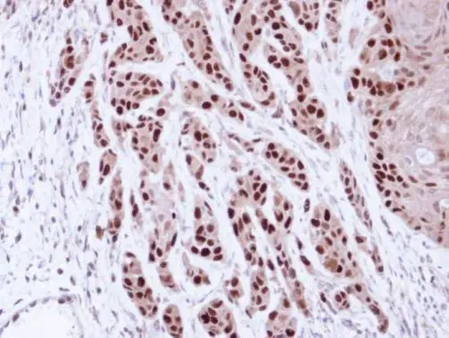
Immunohistochemical analysis of paraffin-embedded Cal27 Xenograft, using MCM7(GTX110278) antibody at 1:100 dilution.
Antigen Retrieval: Trilogy? (EDTA based, pH 8.0) buffer, 15min
 antibody at 1:200 dilution.")

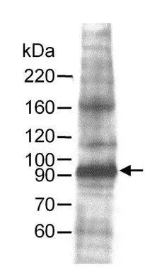
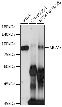
![MCM7 antibody [N2C2], Internal immunoprecipitates MCM7 protein in IP experiments. IP samples: HeLa whole cell extract A. 40 μg HeLa whole cell extract B. Control with 4 μg of preimmune Rabbit IgG C. Immunoprecipitation of MCM7 protein by 4 μg MCM7 antibody [N2C2], Internal (GTX110278) 7.5 % SDS-PAGE The immunoprecipitated MCM7 protein was detected by MCM7 antibody [N2C2], Internal (GTX110278) diluted at 1:1000. [EasyBlot anti-rabbit IgG (GTX221666-01) was used as a secondary reagent]](https://www.genetex.com/upload/website/prouct_img/normal/GTX110278/GTX110278_40051_IP_w_23060500_717.webp "MCM7 antibody [N2C2], Internal immunoprecipitates MCM7 protein in IP experiments. IP samples: HeLa whole cell extract A. 40 μg HeLa whole cell extract B. Control with 4 μg of preimmune Rabbit IgG C. Immunoprecipitation of MCM7 protein by 4 μg MCM7 antibody [N2C2], Internal (GTX110278) 7.5 % SDS-PAGE The immunoprecipitated MCM7 protein was detected by MCM7 antibody [N2C2], Internal (GTX110278) diluted at 1:1000. [EasyBlot anti-rabbit IgG (GTX221666-01) was used as a secondary reagent]")
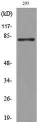
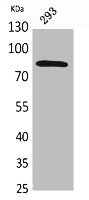
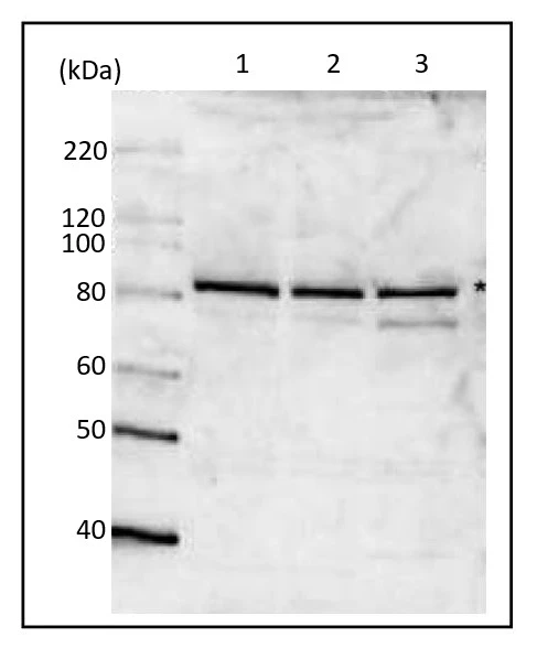
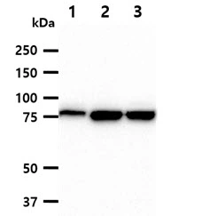
![Various whole cell extracts (30 μg) were separated by 7.5% SDS-PAGE, and the membrane was blotted with MCM7 antibody [N2C2], Internal (GTX110278) diluted at 1:1000. The HRP-conjugated anti-rabbit IgG antibody (GTX213110-01) was used to detect the primary antibody.](https://www.genetex.com/upload/website/prouct_img/normal/GTX110278/GTX110278_40051_20250221_WB_25022521_337.webp "Various whole cell extracts (30 μg) were separated by 7.5% SDS-PAGE, and the membrane was blotted with MCM7 antibody [N2C2], Internal (GTX110278) diluted at 1:1000. The HRP-conjugated anti-rabbit IgG antibody (GTX213110-01) was used to detect the primary antibody.")
![Various whole cell extracts (30 μg) were separated by 7.5% SDS-PAGE, and the membrane was blotted with MCM7 antibody [N2C2], Internal (GTX110278) diluted at 1:1000. The HRP-conjugated anti-rabbit IgG antibody (GTX213110-01) was used to detect the primary antibody.](https://www.genetex.com/upload/website/prouct_img/normal/GTX110278/GTX110278_40051_20250425_WB_M_R_25042920_333.webp "Various whole cell extracts (30 μg) were separated by 7.5% SDS-PAGE, and the membrane was blotted with MCM7 antibody [N2C2], Internal (GTX110278) diluted at 1:1000. The HRP-conjugated anti-rabbit IgG antibody (GTX213110-01) was used to detect the primary antibody.")
![Various whole cell extracts (30 μg) were separated by 7.5% SDS-PAGE, and the membrane was blotted with MCM7 antibody [N2C2], Internal (GTX110278) diluted at 1:1000. The HRP-conjugated anti-rabbit IgG antibody (GTX213110-01) was used to detect the primary antibody.](https://www.genetex.com/upload/website/prouct_img/normal/GTX110278/GTX110278_40051_20250425_WB_M_R_2_25042920_184.webp "Various whole cell extracts (30 μg) were separated by 7.5% SDS-PAGE, and the membrane was blotted with MCM7 antibody [N2C2], Internal (GTX110278) diluted at 1:1000. The HRP-conjugated anti-rabbit IgG antibody (GTX213110-01) was used to detect the primary antibody.")
![Various whole cell extracts (30 μg) were separated by 7.5% SDS-PAGE, and the membrane was blotted with MCM7 antibody [N2C2], Internal (GTX110278) diluted at 1:1000. The HRP-conjugated anti-rabbit IgG antibody (GTX213110-01) was used to detect the primary antibody.](https://www.genetex.com/upload/website/prouct_img/normal/GTX110278/GTX110278_40051_20250425_WB_25042920_743.webp "Various whole cell extracts (30 μg) were separated by 7.5% SDS-PAGE, and the membrane was blotted with MCM7 antibody [N2C2], Internal (GTX110278) diluted at 1:1000. The HRP-conjugated anti-rabbit IgG antibody (GTX213110-01) was used to detect the primary antibody.")
Immunohistochemical analysis of paraffin-embedded Cal27 Xenograft, using MCM7(GTX110278) antibody at 1:100 dilution.
Antigen Retrieval: Trilogy? (EDTA based, pH 8.0) buffer, 15min
MCM7 antibody [N2C2], Internal
GTX110278
ApplicationsImmunoFluorescence, ImmunoPrecipitation, Western Blot, ChIP Chromatin ImmunoPrecipitation, ImmunoCytoChemistry, ImmunoHistoChemistry, ImmunoHistoChemistry Paraffin
Product group Antibodies
ReactivityHuman
TargetMCM7
Overview
- SupplierGeneTex
- Product NameMCM7 antibody [N2C2], Internal
- Delivery Days Customer9
- Application Supplier NoteWB: 1:500-1:3000. ICC/IF: 1:100-1:1000. IHC-P: 1:100-1:1000. IP: 1:100-1:500. *Optimal dilutions/concentrations should be determined by the researcher.Not tested in other applications.
- ApplicationsImmunoFluorescence, ImmunoPrecipitation, Western Blot, ChIP Chromatin ImmunoPrecipitation, ImmunoCytoChemistry, ImmunoHistoChemistry, ImmunoHistoChemistry Paraffin
- CertificationResearch Use Only
- ClonalityPolyclonal
- Concentration0.76 mg/ml
- ConjugateUnconjugated
- Gene ID4176
- Target nameMCM7
- Target descriptionminichromosome maintenance complex component 7
- Target synonymsCDC47, MCM2, P1.1-MCM3, P1CDC47, P85MCM, PNAS146, PPP1R104, DNA replication licensing factor MCM7, CDC47 homolog, homolog of S. cerevisiae Cdc47, minichromosome maintenance deficient 7, protein phosphatase 1, regulatory subunit 104
- HostRabbit
- IsotypeIgG
- Protein IDP33993
- Protein NameDNA replication licensing factor MCM7
- Scientific DescriptionThe protein encoded by this gene is one of the highly conserved mini-chromosome maintenance proteins (MCM) that are essential for the initiation of eukaryotic genome replication. The hexameric protein complex formed by the MCM proteins is a key component of the pre-replication complex (pre_RC) and may be involved in the formation of replication forks and in the recruitment of other DNA replication related proteins. The MCM complex consisting of this protein and MCM2, 4 and 6 proteins possesses DNA helicase activity, and may act as a DNA unwinding enzyme. Cyclin D1-dependent kinase, CDK4, is found to associate with this protein, and may regulate the binding of this protein with the tumorsuppressor protein RB1/RB. Alternatively spliced transcript variants encoding distinct isoforms have been reported. [provided by RefSeq]
- ReactivityHuman
- Storage Instruction-20°C or -80°C,2°C to 8°C
- UNSPSC41116161
Datasheet
Related products
Product group Antibodies
Anti-MCM7 AntibodyA98047
ApplicationsWestern Blot, ELISA
ReactivityHuman, Mouse, Rat
- SizePrice
Product group Antibodies
Anti-MCM7 Antibody144-01138
ApplicationsImmunoFluorescence, Western Blot
ReactivityHuman, Mouse
TargetMCM7
- SizePrice
Product group Antibodies
ApplicationsFlow Cytometry, Western Blot, ImmunoCytoChemistry
ReactivityHuman, Rat
TargetMCM7
- SizePrice
Product group Antibodies
MCM7 AntibodyCSB-PA005604
ApplicationsWestern Blot, ELISA
ReactivityHuman, Mouse, Rat
TargetMCM7
- SizePrice
Product group Antibodies
MCM7 antibodyGTX23732
ApplicationsWestern Blot, Other Application
ReactivityHuman, Mouse
TargetMCM7
- SizePrice
Product group Antibodies
MCM7 antibodyGTX00894
ApplicationsFlow Cytometry, ImmunoFluorescence, ImmunoPrecipitation, Western Blot, ChIP Chromatin ImmunoPrecipitation, ImmunoCytoChemistry
ReactivityHamster, Human, Mouse, Rat
TargetMCM7
- SizePrice
![IHC-P analysis of human lymph node tissue using GTX04444 MCM7 antibody [MSVA-507R] HistoMAX?. In lymph nodes a particularly strong MCM7 staining occurs in most cells of germinal centres.](https://www.genetex.com/upload/website/prouct_img/normal/GTX04444/GTX04444_20230728_IHC-P_74_23072722_267.webp)
Product group Antibodies
ApplicationsImmunoHistoChemistry, ImmunoHistoChemistry Paraffin
ReactivityHuman
TargetMCM7
- SizePrice

Product group Antibodies
Anti-MCM7 AntibodyHPA003898
ApplicationsWestern Blot, ImmunoCytoChemistry, ImmunoHistoChemistry
ReactivityHuman, Mouse, Rat
TargetMCM7
- SizePrice
Product group Antibodies
MCM7 antibodyGTX54354
ApplicationsImmunoFluorescence, ImmunoPrecipitation, Western Blot, ImmunoCytoChemistry
ReactivityHuman
TargetMCM7
- SizePrice
Product group Antibodies
MCM7 antibody [AT118H7]GTX57715
ApplicationsImmunoFluorescence, Western Blot, ImmunoCytoChemistry
ReactivityHuman
TargetMCM7
- SizePrice