

MDH1 Monoclonal Antibody
BSM-60336M
ApplicationsImmunoFluorescence, Western Blot, ImmunoCytoChemistry
Product group Antibodies
TargetMDH1
Overview
- SupplierBioss Antibodies
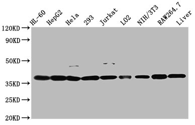
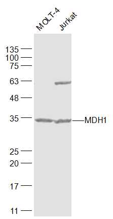
- Product NameMDH1 Monoclonal Antibody
- Delivery Days Customer16
- ApplicationsImmunoFluorescence, Western Blot, ImmunoCytoChemistry
- Applications SupplierWB(1:300-5000), ICC/IF(1:50-100)
- CertificationResearch Use Only
- ClonalityMonoclonal
- Clone IDB10D3
- Concentration1 mg/ml
- ConjugateUnconjugated
- Gene ID4190
- Target nameMDH1
- Target descriptionmalate dehydrogenase 1
- Target synonymscytosolic malate dehydrogenase; DEE88; diiodophenylpyruvate reductase; EIEE88; epididymis secretory protein Li 32; HEL-S-32; KAR; malate dehydrogenase 1, NAD (soluble); malate dehydrogenase, cytoplasmic; malate dehydrogenase, peroxisomal; MDHA; MDH-s; MGC:1375; MOR2; soluble malate dehydrogenase
- HostMouse
- IsotypeIgG1
- Protein IDP40925
- Protein NameMalate dehydrogenase, cytoplasmic
- Storage Instruction-20°C
- UNSPSC12352203
Related products
Product group Antibodies

Goat anti-MDH1 / MOR2EB11994
ApplicationsWestern Blot, ELISA
TargetMDH1
- SizePrice
Product group Antibodies
MDH1 Polyclonal AntibodyCAC15653
ApplicationsImmunoFluorescence, Western Blot, ELISA, ImmunoHistoChemistry
TargetMDH1
- SizePrice
Product group Antibodies
MDH1 AntibodyCSB-PA013621EA01HU
ApplicationsImmunoFluorescence, Western Blot, ELISA, ImmunoHistoChemistry
TargetMDH1
- SizePrice

Product group Antibodies
Anti-MDH1 Antibody Picoband(r)A04262-2-CARRIER-FREE
ApplicationsFlow Cytometry, Western Blot, ELISA
TargetMDH1
- SizePrice
Product group Antibodies
References
MDH1 Polyclonal AntibodyBS-3996R
ApplicationsFlow Cytometry, ImmunoFluorescence, Western Blot, ELISA, ImmunoCytoChemistry, ImmunoHistoChemistry, ImmunoHistoChemistry Frozen, ImmunoHistoChemistry Paraffin
TargetMDH1
- SizePrice