

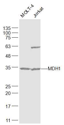
Lane 1: MOLT-4 cell lysates; Lane 2: Jurkat cell lysates probed with MDH1 Polyclonal Antibody, Unconjugated (bs-3996R) at 1:1000 dilution and 4˚C overnight incubation. Followed by conjugated secondary antibody incubation at 1:20000 for 60 min at 37˚C.
 at 1:1000 dilution and 4˚C overnight incubation. Followed by conjugated secondary antibody incubation at 1:20000 for 60 min at 37˚C.")
 at 1:200 followed by conjugation to the secondary antibody and DAB staining")
Lane 1: MOLT-4 cell lysates; Lane 2: Jurkat cell lysates probed with MDH1 Polyclonal Antibody, Unconjugated (bs-3996R) at 1:1000 dilution and 4˚C overnight incubation. Followed by conjugated secondary antibody incubation at 1:20000 for 60 min at 37˚C.
MDH1 Polyclonal Antibody
BS-3996R
ApplicationsFlow Cytometry, ImmunoFluorescence, Western Blot, ELISA, ImmunoCytoChemistry, ImmunoHistoChemistry, ImmunoHistoChemistry Frozen, ImmunoHistoChemistry Paraffin
Product group Antibodies
ReactivityBovine, Canine, Chicken, Equine, Human, Mouse, Porcine, Rabbit, Rat
TargetMDH1
Overview
- SupplierBioss
- Product NameMDH1 Polyclonal Antibody
- Delivery Days Customer16
- ApplicationsFlow Cytometry, ImmunoFluorescence, Western Blot, ELISA, ImmunoCytoChemistry, ImmunoHistoChemistry, ImmunoHistoChemistry Frozen, ImmunoHistoChemistry Paraffin
- Applications SupplierWB(1:300-5000), ELISA(1:500-1000), FCM(1:20-100), IHC-P(1:200-400), IHC-F(1:100-500), IF(IHC-P)(1:50-200), IF(IHC-F)(1:50-200), IF(ICC)(1:50-200)
- CertificationResearch Use Only
- ClonalityPolyclonal
- Concentration1 ug/ul
- ConjugateUnconjugated
- Gene ID4190
- Target nameMDH1
- Target descriptionmalate dehydrogenase 1
- Target synonymsDEE88, EIEE88, HEL-S-32, KAR, MDH-s, MDHA, MGC:1375, MOR2, malate dehydrogenase, cytoplasmic, malate dehydrogenase, peroxisomal, aromatic alpha-keto acid reductase, cytosolic malate dehydrogenase, diiodophenylpyruvate reductase, epididymis secretory protein Li 32, malate dehydrogenase 1, NAD (soluble), soluble malate dehydrogenase
- HostRabbit
- IsotypeIgG
- Protein IDP40925
- Protein NameMalate dehydrogenase, cytoplasmic
- ReactivityBovine, Canine, Chicken, Equine, Human, Mouse, Porcine, Rabbit, Rat
- Storage Instruction-20°C
- UNSPSC41116161
References
- STAT3-RXR-Nrf2 activates systemic redox and energy homeostasis upon steep decline in pO2 gradient. Paul S et al., 2018 Apr, Redox BiolRead this paper
Datasheet
Related products
Product group Antibodies
Anti-MDH1 AntibodyA116675
ApplicationsDot Blot, ImmunoFluorescence, Western Blot, ELISA
ReactivityHuman
- SizePrice

Product group Antibodies
Anti-MDH1 Antibody Picoband(r)A04262-2-CARRIER-FREE
ApplicationsFlow Cytometry, Western Blot, ELISA
ReactivityHuman, Mouse, Rat
TargetMDH1
- SizePrice
Product group Antibodies
Anti-MDH1 Antibody144-07563
ApplicationsWestern Blot
ReactivityHuman, Mouse, Rat
TargetMDH1
- SizePrice
Product group Antibodies
MDH1 AntibodyCSB-PA013621EA01HU
ApplicationsImmunoFluorescence, Western Blot, ELISA, ImmunoHistoChemistry
ReactivityHuman, Mouse
TargetMDH1
- SizePrice
Product group Antibodies

Goat anti-MDH1 / MOR2EB11994
ApplicationsWestern Blot, ELISA
ReactivityBovine, Canine, Human, Mouse, Porcine, Rat
TargetMDH1
- SizePrice
Product group Antibodies
MDH1 Polyclonal AntibodyCAC15653
ApplicationsImmunoFluorescence, Western Blot, ELISA, ImmunoHistoChemistry
ReactivityMouse
TargetMDH1
- SizePrice

Product group Antibodies

MDH1 AntibodyLS-C401833
ApplicationsWestern Blot, ELISA
ReactivityHuman, Mouse, Rat
TargetMDH1
- SizePrice

Product group Antibodies
Anti-MDH1 AntibodyHPA027296
ApplicationsWestern Blot, ImmunoCytoChemistry, ImmunoHistoChemistry
ReactivityHuman
TargetMDH1
- SizePrice