


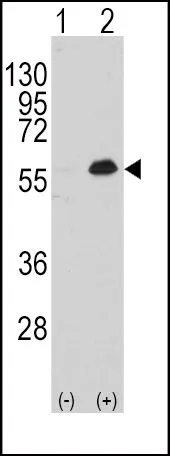
Various whole cell extracts (30 μg) were separated by 10% SDS-PAGE, and the membrane was blotted with MEF2C antibody [HL2811] (GTX639952) diluted at 1:1000. The HRP-conjugated anti-rabbit IgG antibody (GTX213110-01) was used to detect the primary antibody.
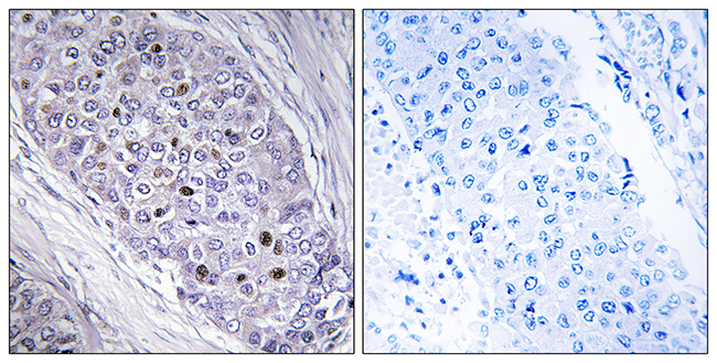
![MEF2C antibody [HL2811] detects MEF2C protein at nucleus by immunohistochemical analysis. Sample: Paraffin-embedded rat heart. MEF2C stained by MEF2C antibody [HL2811] (GTX639952) diluted at 1:100. Antigen Retrieval: Citrate buffer, pH 6.0, 15 min](https://www.genetex.com/upload/website/prouct_img/normal/GTX639952/GTX639952_T-45348_20240401_IHC-P_R_24041019_583.webp "MEF2C antibody [HL2811] detects MEF2C protein at nucleus by immunohistochemical analysis. Sample: Paraffin-embedded rat heart. MEF2C stained by MEF2C antibody [HL2811] (GTX639952) diluted at 1:100. Antigen Retrieval: Citrate buffer, pH 6.0, 15 min")
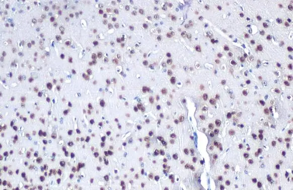
![MEF2C antibody [HL2811] detects MEF2C protein at nucleus by immunohistochemical analysis. Sample: Paraffin-embedded mouse brain. MEF2C stained by MEF2C antibody [HL2811] (GTX639952) diluted at 1:100. Antigen Retrieval: Citrate buffer, pH 6.0, 15 min](https://www.genetex.com/upload/website/prouct_img/normal/GTX639952/GTX639952_T-45348_20240401_IHC-P_M_24041019_456.webp "MEF2C antibody [HL2811] detects MEF2C protein at nucleus by immunohistochemical analysis. Sample: Paraffin-embedded mouse brain. MEF2C stained by MEF2C antibody [HL2811] (GTX639952) diluted at 1:100. Antigen Retrieval: Citrate buffer, pH 6.0, 15 min")
![MEF2C antibody [HL2811] detects MEF2C protein at nucleus by immunofluorescent analysis. Sample: HeLa cells were fixed in 4% paraformaldehyde at RT for 15 min. Green: MEF2C stained by MEF2C antibody [HL2811] (GTX639952) diluted at 1:500. Red: alpha Tubulin, a cytoskeleton marker, stained by alpha Tubulin antibody [GT114] (GTX628802) diluted at 1:1000.](https://www.genetex.com/upload/website/prouct_img/normal/GTX639952/GTX639952_T-45348_20240329_ICC_IF_24041019_782.webp "MEF2C antibody [HL2811] detects MEF2C protein at nucleus by immunofluorescent analysis. Sample: HeLa cells were fixed in 4% paraformaldehyde at RT for 15 min. Green: MEF2C stained by MEF2C antibody [HL2811] (GTX639952) diluted at 1:500. Red: alpha Tubulin, a cytoskeleton marker, stained by alpha Tubulin antibody [GT114] (GTX628802) diluted at 1:1000.")
![Various whole cell extracts (30 μg) were separated by 10% SDS-PAGE, and the membrane was blotted with MEF2C antibody [HL2811] (GTX639952) diluted at 1:1000. The HRP-conjugated anti-rabbit IgG antibody (GTX213110-01) was used to detect the primary antibody. Corresponding RNA expression data for the same cell lines are based on Human Protein Atlas program.](https://www.genetex.com/upload/website/prouct_img/normal/GTX639952/GTX639952_45418_20240524_WB_TPM_watermark_24052802_757.webp "Various whole cell extracts (30 μg) were separated by 10% SDS-PAGE, and the membrane was blotted with MEF2C antibody [HL2811] (GTX639952) diluted at 1:1000. The HRP-conjugated anti-rabbit IgG antibody (GTX213110-01) was used to detect the primary antibody. Corresponding RNA expression data for the same cell lines are based on Human Protein Atlas program.")
Various whole cell extracts (30 μg) were separated by 10% SDS-PAGE, and the membrane was blotted with MEF2C antibody [HL2811] (GTX639952) diluted at 1:1000. The HRP-conjugated anti-rabbit IgG antibody (GTX213110-01) was used to detect the primary antibody.
MEF2C antibody [HL2811]
GTX639952
ApplicationsImmunoFluorescence, Western Blot, ImmunoCytoChemistry, ImmunoHistoChemistry, ImmunoHistoChemistry Paraffin
Product group Antibodies
ReactivityHuman, Mouse, Rat
TargetMEF2C
Overview
- SupplierGeneTex
- Product NameMEF2C antibody [HL2811]
- Delivery Days Customer7
- Application Supplier NoteWB: 1:500-1:3000. *Optimal dilutions/concentrations should be determined by the researcher.Not tested in other applications.
- ApplicationsImmunoFluorescence, Western Blot, ImmunoCytoChemistry, ImmunoHistoChemistry, ImmunoHistoChemistry Paraffin
- CertificationResearch Use Only
- ClonalityMonoclonal
- Clone IDHL2811
- Concentration1 mg/ml
- ConjugateUnconjugated
- Gene ID4208
- Target nameMEF2C
- Target descriptionmyocyte enhancer factor 2C
- Target synonymsC5DELq14.3, DEL5q14.3, NEDHSIL, myocyte-specific enhancer factor 2C, MADS box transcription enhancer factor 2, polypeptide C
- HostRabbit
- IsotypeIgG
- Protein IDQ06413
- Protein NameMyocyte-specific enhancer factor 2C
- Scientific DescriptionThis locus encodes a member of the MADS box transcription enhancer factor 2 (MEF2) family of proteins, which play a role in myogenesis. The encoded protein, MEF2 polypeptide C, has both trans-activating and DNA binding activities. This protein may play a role in maintaining the differentiated state of muscle cells. Mutations and deletions at this locus have been associated with severe cognitive disability, stereotypic movements, epilepsy, and cerebral malformation. Alternatively spliced transcript variants have been described. [provided by RefSeq, Jul 2010]
- ReactivityHuman, Mouse, Rat
- Storage Instruction-20°C or -80°C,2°C to 8°C
- UNSPSC41116161
Datasheet
Related products
Product group Antibodies

MEF2C (Phospho-Ser396) AntibodyABX012699
ApplicationsWestern Blot, ELISA, ImmunoHistoChemistry
- SizePrice

Product group Antibodies
Anti-MEF2C Antibody Picoband(r)A01131-1-CARRIER-FREE
ApplicationsFlow Cytometry, Western Blot, ImmunoHistoChemistry
ReactivityHuman, Mouse, Rat
TargetMEF2C
- SizePrice

Product group Antibodies
Anti-MEF2C AntibodyAMAB90727
ApplicationsWestern Blot, ImmunoCytoChemistry, ImmunoHistoChemistry
ReactivityHuman
TargetMEF2C
- SizePrice
Product group Antibodies

Anti-MFE2C [AbAb-MFE2C]AB04171-1.1
ApplicationsImmunoFluorescence, Western Blot, ELISA, Other Application
ReactivityHuman
TargetMEF2C
- SizePrice
Product group Antibodies
Anti-MEF2C AntibodyA98862
ApplicationsELISA, ImmunoHistoChemistry
ReactivityHuman, Mouse
- SizePrice
Product group Antibodies

MEF2C AntibodyLS-C763475
ApplicationsImmunoHistoChemistry
ReactivityHuman
TargetMEF2C
- SizePrice
Product group Antibodies
Phospho-MEF2C (S396) AntibodyCSB-PA010169
ApplicationsWestern Blot, ELISA, ImmunoHistoChemistry
ReactivityHuman, Mouse
TargetMEF2C
- SizePrice
Product group Antibodies
MEF2C antibodyGTX81913
ApplicationsWestern Blot, ImmunoHistoChemistry, ImmunoHistoChemistry Paraffin
ReactivityHuman
TargetMEF2C
- SizePrice
Product group Antibodies
MEF2C antibodyGTX105433
ApplicationsImmunoFluorescence, ImmunoPrecipitation, Western Blot, ImmunoCytoChemistry, ImmunoHistoChemistry, ImmunoHistoChemistry Frozen, ImmunoHistoChemistry Paraffin
ReactivityHuman, Mouse
TargetMEF2C
- SizePrice
![MEF2C antibody detects MEF2C protein by immunofluorescent analysis. Sample: DIV10 rat E18 primary cortical neuron cells were fixed in 4% paraformaldehyde at RT for 15 min. Green: MEF2C stained by MEF2C antibody (GTX111134) diluted at 1:500. Red: beta Tubulin 3/ Tuj1, stained by beta Tubulin 3/ Tuj1 antibody [GT11710] (GTX631836) diluted at 1:500. Blue: Fluoroshield with DAPI (GTX30920).](https://www.genetex.com/upload/website/prouct_img/normal/GTX111134/GTX111134_40464_20181227_ICC_IF_R_w_23060500_709.webp)
Product group Antibodies
MEF2C antibodyGTX111134
ApplicationsImmunoFluorescence, Western Blot, ImmunoCytoChemistry, ImmunoHistoChemistry, ImmunoHistoChemistry Frozen
ReactivityHuman, Mouse, Rat
TargetMEF2C
- SizePrice