

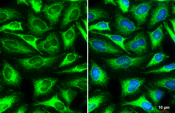
MFF antibody detects MFF protein at mitochondria by immunofluorescent analysis. Sample: HeLa cells were fixed in ice-cold MeOH for 5 min. Green: MFF stained by MFF antibody (GTX135140) diluted at 1:500. Blue: Fluoroshield with DAPI (GTX30920). Scale bar= 10 μm.
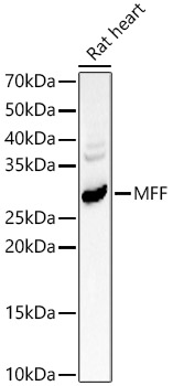
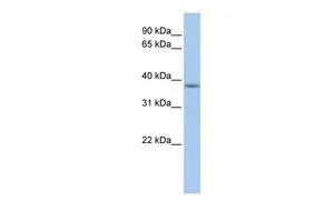
 were separated by 10% SDS-PAGE, and the membrane was blotted with MFF antibody (GTX135140) diluted at 1:1000. The HRP-conjugated anti-rabbit IgG antibody (GTX213110-01) was used to detect the primary antibody.")
MFF antibody detects MFF protein at mitochondria by immunofluorescent analysis. Sample: HeLa cells were fixed in ice-cold MeOH for 5 min. Green: MFF stained by MFF antibody (GTX135140) diluted at 1:500. Blue: Fluoroshield with DAPI (GTX30920). Scale bar= 10 μm.
MFF antibody
GTX135140
ApplicationsImmunoFluorescence, Western Blot, ImmunoCytoChemistry
Product group Antibodies
ReactivityHuman
TargetMFF
Overview
- SupplierGeneTex
- Product NameMFF antibody
- Delivery Days Customer9
- Application Supplier NoteWB: 1:1000. *Optimal dilutions/concentrations should be determined by the researcher.Not tested in other applications.
- ApplicationsImmunoFluorescence, Western Blot, ImmunoCytoChemistry
- CertificationResearch Use Only
- ClonalityPolyclonal
- Concentration1.41 mg/ml
- ConjugateUnconjugated
- Gene ID56947
- Target nameMFF
- Target descriptionmitochondrial fission factor
- Target synonymsC2orf33, EMPF2, GL004, mitochondrial fission factor
- HostRabbit
- IsotypeIgG
- Protein IDQ9GZY8
- Protein NameMitochondrial fission factor
- Scientific DescriptionThis is a nuclear gene encoding a protein that functions in mitochondrial and peroxisomal fission. The encoded protein recruits dynamin-1-like protein (DNM1L) to mitochondria. There are multiple pseudogenes for this gene on chromosomes 1, 5, and X. Alternative splicing results in multiple transcript variants. [provided by RefSeq, Mar 2013]
- ReactivityHuman
- Storage Instruction-20°C,2°C to 8°C
- UNSPSC41116161
Datasheet
Related products

Product group Antibodies
Anti-MFF Antibody Picoband(r)A02563-1-CARRIER-FREE
ApplicationsFlow Cytometry, ImmunoFluorescence, Western Blot, ELISA, ImmunoCytoChemistry, ImmunoHistoChemistry
ReactivityHuman, Mouse, Rat
TargetMFF
- SizePrice
Product group Antibodies
Anti-MFF Antibody144-65840
ApplicationsWestern Blot, ImmunoHistoChemistry
ReactivityHuman
TargetMFF
- SizePrice
Product group Antibodies

ApplicationsWestern Blot, ELISA
- SizePrice
Product group Antibodies
Anti-MFF AntibodyA14609
ApplicationsWestern Blot, ImmunoHistoChemistry
ReactivityHuman, Mouse, Rat
- SizePrice
Product group Antibodies

MFF AntibodyLS-C747496
ApplicationsWestern Blot
ReactivityHuman
TargetMFF
- SizePrice
Product group Antibodies
MFF Polyclonal AntibodyBS-7628R
ApplicationsImmunoFluorescence, Western Blot, ELISA, ImmunoCytoChemistry, ImmunoHistoChemistry, ImmunoHistoChemistry Frozen, ImmunoHistoChemistry Paraffin
ReactivityBovine, Chicken, Equine, Human, Mouse, Porcine, Rabbit, Rat, Sheep, Zebra Fish
- SizePrice
Product group Antibodies
MFF AntibodyCSB-PA013751LA01HU
ApplicationsWestern Blot, ELISA
ReactivityHuman
TargetMFF
- SizePrice
Product group Antibodies
MFF Polyclonal AntibodyCAC15837
ApplicationsWestern Blot, ELISA
TargetMFF
- SizePrice
Product group Antibodies
Anti-MFF AntibodyHPA010968
ApplicationsImmunoHistoChemistry
ReactivityHuman
TargetMFF
- SizePrice
Product group Antibodies
MFF antibody, C-termGTX46061
ApplicationsWestern Blot
ReactivityHuman
TargetMFF
- SizePrice