

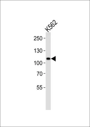
WB analysis of K562 cell lysate (35ug/lane) using GTX53672 MIB1 antibody.

WB analysis of K562 cell lysate (35ug/lane) using GTX53672 MIB1 antibody.
MIB1 antibody
GTX53672
ApplicationsImmunoFluorescence, Western Blot, ImmunoCytoChemistry, ImmunoHistoChemistry, ImmunoHistoChemistry Paraffin
Product group Antibodies
ReactivityHuman
TargetMIB1
Overview
- SupplierGeneTex
- Product NameMIB1 antibody
- Delivery Days Customer9
- Application Supplier NoteWB: 1:1000. ICC/IF: 1:20-1:100. IHC-P: 1:50-1:100. *Optimal dilutions/concentrations should be determined by the researcher.Not tested in other applications.
- ApplicationsImmunoFluorescence, Western Blot, ImmunoCytoChemistry, ImmunoHistoChemistry, ImmunoHistoChemistry Paraffin
- CertificationResearch Use Only
- ClonalityPolyclonal
- ConjugateUnconjugated
- Gene ID57534
- Target nameMIB1
- Target descriptionMIB E3 ubiquitin protein ligase 1
- Target synonymsDIP-1, DIP1, LVNC7, MIB, ZZANK2, ZZZ6, E3 ubiquitin-protein ligase MIB1, DAPK-interacting protein 1, RING-type E3 ubiquitin transferase MIB1, mindbomb E3 ubiquitin protein ligase 1, ubiquitin ligase mind bomb, zinc finger ZZ type with ankyrin repeat domain protein 2
- HostRabbit
- IsotypeIgG
- Protein IDQ86YT6
- Protein NameE3 ubiquitin-protein ligase MIB1
- Scientific DescriptionThis gene encodes a protein containing multiple ankyrin repeats and RING finger domains that functions as an E3 ubiquitin ligase. The encoded protein positively regulates Notch signaling by ubiquitinating the Notch receptors, thereby facilitating their endocytosis. This protein may also promote the ubiquitination and degradation of death-associated protein kinase 1 (DAPK1). [provided by RefSeq, Jun 2013]
- ReactivityHuman
- Storage Instruction-20°C or -80°C,2°C to 8°C
- UNSPSC41116161
Datasheet
Related products

Product group Antibodies
Anti-Mib1/Mindbomb Antibody Picoband(r)A01387-1-CARRIER-FREE
ApplicationsWestern Blot, ELISA
ReactivityHuman, Mouse, Rat
TargetMIB1
- SizePrice
Product group Antibodies
Anti-MIB1 Antibody144-08588
ApplicationsWestern Blot
ReactivityHuman, Mouse, Rat
TargetMIB1
- SizePrice
Product group Antibodies
Anti-Mib1/Mindbomb AntibodyA326263
ApplicationsWestern Blot, ELISA, ImmunoHistoChemistry
ReactivityHuman
- SizePrice
Product group Antibodies
MIB1 Recombinant AntibodyBSM-61914R
ApplicationsImmunoFluorescence, Western Blot, ImmunoCytoChemistry
ReactivityHuman, Mouse
TargetMIB1
- SizePrice
Product group Antibodies
MIB1 AntibodyCSB-PA13289A0RB
ApplicationsImmunoFluorescence, Western Blot, ELISA, ImmunoHistoChemistry
ReactivityHuman
TargetMIB1
- SizePrice
Product group Antibodies

Goat anti-Mindbomb 1 / MIB1EB11532
ApplicationsWestern Blot, ELISA, ImmunoHistoChemistry
ReactivityBovine, Canine, Human, Mouse, Rat
TargetMIB1
- SizePrice
Product group Antibodies
MIB1 Polyclonal AntibodyCAC14135
ApplicationsImmunoFluorescence, Western Blot, ELISA, ImmunoHistoChemistry
TargetMIB1
- SizePrice
Product group Antibodies

MIB1 AntibodyLS-C410123
ApplicationsWestern Blot
ReactivityHuman, Mouse
TargetMIB1
- SizePrice
Product group Antibodies
MIB1 antibodyGTX31573
ApplicationsImmunoFluorescence, Western Blot, ELISA, ImmunoCytoChemistry
ReactivityHuman, Mouse, Rat
TargetMIB1
- SizePrice