

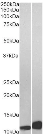
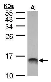
WB analysis of Daudi (A) and Jurkat (B) lysate using GTX89535 MIF antibody, C-term. Dilution : 0.01μg/ml Loading : 35μg protein in RIPA buffer


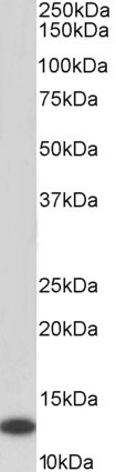
 Detection : GTX89535 (1.5μg/ml)")
WB analysis of Daudi (A) and Jurkat (B) lysate using GTX89535 MIF antibody, C-term. Dilution : 0.01μg/ml Loading : 35μg protein in RIPA buffer
MIF antibody, C-term
GTX89535
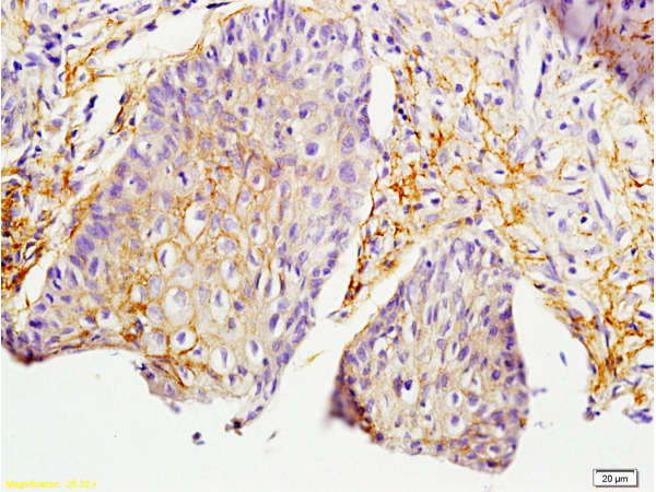
ApplicationsWestern Blot, ELISA, ImmunoHistoChemistry, ImmunoHistoChemistry Paraffin
Product group Antibodies
ReactivityHuman
TargetMIF
Overview
- SupplierGeneTex
- Product NameMIF antibody, C-term
- Delivery Days Customer7
- Application Supplier NoteWB: 0.01-0.03microg/ml. IHC-P: 5microg/ml. *Optimal dilutions/concentrations should be determined by the researcher.Not tested in other applications.
- ApplicationsWestern Blot, ELISA, ImmunoHistoChemistry, ImmunoHistoChemistry Paraffin
- CertificationResearch Use Only
- ClonalityPolyclonal
- Concentration0.50 mg/ml
- ConjugateUnconjugated
- Gene ID4282
- Target nameMIF
- Target descriptionmacrophage migration inhibitory factor
- Target synonymsGIF, GLIF, MMIF, macrophage migration inhibitory factor, L-dopachrome isomerase, L-dopachrome tautomerase, epididymis secretory sperm binding protein, macrophage migration inhibitory factor (glycosylation-inhibiting factor), phenylpyruvate tautomerase
- HostGoat
- IsotypeIgG
- Protein IDP14174
- Protein NameMacrophage migration inhibitory factor
- Scientific DescriptionThis gene encodes a lymphokine involved in cell-mediated immunity, immunoregulation, and inflammation. It plays a role in the regulation of macrophage function in host defense through the suppression of anti-inflammatory effects of glucocorticoids. This lymphokine and the JAB1 protein form a complex in the cytosol near the peripheral plasma membrane, which may indicate an additional role in integrin signaling pathways. [provided by RefSeq, Jul 2008]
- ReactivityHuman
- Storage Instruction-20°C or -80°C,2°C to 8°C
- UNSPSC41116161
Datasheet
Related products
Product group Antibodies
ApplicationsWestern Blot, ELISA
ReactivityHuman
- SizePrice
Product group Antibodies
Anti-MIF Antibody130-10009
ApplicationsWestern Blot, ELISA
ReactivityHuman
- SizePrice
Product group Antibodies

Kininogen 1 (KNG1) AntibodyABX109959
ApplicationsImmunoFluorescence, Western Blot, ELISA, ImmunoCytoChemistry, ImmunoHistoChemistry
- SizePrice
Product group Antibodies
References
MIF Polyclonal AntibodyBS-1044R
ApplicationsFlow Cytometry, ImmunoFluorescence, Western Blot, ELISA, ImmunoCytoChemistry, ImmunoHistoChemistry, ImmunoHistoChemistry Frozen, ImmunoHistoChemistry Paraffin
TargetMIF
- SizePrice
Product group Antibodies
MIF AntibodyCSB-PA003240
ApplicationsWestern Blot, ELISA
ReactivityHuman, Mouse, Rat
TargetMIF
- SizePrice
Product group Antibodies

Goat anti-MIF, BiotinylatedEB06765-B
ApplicationsWestern Blot, ELISA
ReactivityHuman
TargetMIF
- SizePrice
Product group Antibodies
Mif Polyclonal AntibodyCAC07247
ApplicationsImmunoFluorescence, Western Blot, ELISA, ImmunoHistoChemistry
ReactivityMouse, Rat
TargetMIF
- SizePrice
Product group Antibodies
MIF antibody [2Ar3]GTX14575
ApplicationsWestern Blot
ReactivityHuman
TargetMIF
- SizePrice
Product group Antibodies

MIF Antibody (Preservative Free)LS-C149173
ApplicationsWestern Blot, ELISA
ReactivityHuman
TargetMIF
- SizePrice
Product group Antibodies
MIF antibody [N1C3]GTX101162
ApplicationsImmunoFluorescence, Western Blot, ELISA, ImmunoCytoChemistry, ImmunoHistoChemistry, ImmunoHistoChemistry Frozen, ImmunoHistoChemistry Paraffin
ReactivityHuman, Mouse, Rat
TargetMIF
- SizePrice