

Mitochondrial Fission 1 Protein Antibody (YA2098)
HY-P82353
TargetFIS1
Product group Antibodies
Overview
- SupplierMedChem Express
- Product NameMitochondrial Fission 1 Protein Antibody (YA2098)
- Delivery Days Customer5
- CertificationResearch Use Only
- ClonalityMonoclonal
- Gene ID51024
- Target nameFIS1
- Target descriptionfission, mitochondrial 1
- Target synonymsCGI-135; FIS1 homolog; fission 1 (mitochondrial outer membrane) homolog; H_NH0132A01.6; hFis1; mitochondrial fission 1 protein; mitochondrial fission molecule; tetratricopeptide repeat domain 11; tetratricopeptide repeat protein 11; TPR repeat protein 11; TTC11
- HostRabbit
- IsotypeIgG
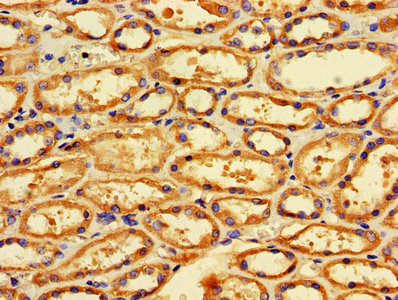
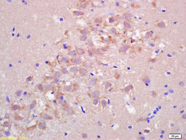
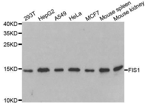
- Scientific DescriptionMitochondrial Fission 1 Protein Antibody (YA2098) is a rabbit-derived non-conjugated IgG antibody (Clone NO.: YA2098), targeting Mitochondrial Fission 1 Protein, with a predicted molecular weight of 17 kDa (observed band size: 17 kDa). Mitochondrial Fission 1 Protein Antibody (YA2098) can be used for WB, IHC-P, ICC/IF experiment in human, cow background.
- Storage Instruction-20°C
- UNSPSC12352203
Related products
Product group Antibodies
Fis1 Polyclonal AntibodyCAC07594
ApplicationsImmunoFluorescence, ELISA, ImmunoHistoChemistry
TargetFIS1
- SizePrice
Product group Antibodies
FIS1 AntibodyCSB-PA008684LA01HU
ApplicationsImmunoFluorescence, ELISA, ImmunoHistoChemistry
TargetFIS1
- SizePrice
Product group Antibodies
References
FIS1 antibodyGTX111010
ApplicationsImmunoFluorescence, Western Blot, ImmunoCytoChemistry, ImmunoHistoChemistry, ImmunoHistoChemistry Paraffin
TargetFIS1
- SizePrice

Product group Antibodies
Anti-FIS1 AntibodyHPA017430
ApplicationsWestern Blot, ImmunoCytoChemistry, ImmunoHistoChemistry
TargetFIS1
- SizePrice
Product group Antibodies
Anti-FIS1 (N-term) Antibody102-23904
ApplicationsWestern Blot
TargetFIS1
- SizePrice
Product group Antibodies
References
FIS1 Polyclonal AntibodyBS-7646R
ApplicationsImmunoFluorescence, Western Blot, ELISA, ImmunoCytoChemistry, ImmunoHistoChemistry, ImmunoHistoChemistry Frozen, ImmunoHistoChemistry Paraffin
TargetFIS1
- SizePrice
Product group Antibodies

FIS1 AntibodyLS-C401687
ApplicationsWestern Blot, ELISA, ImmunoHistoChemistry
TargetFIS1
- SizePrice
Product group Antibodies
Anti-FIS1 AntibodyA31072
ApplicationsImmunoFluorescence, Western Blot, ImmunoHistoChemistry
- SizePrice

Product group Antibodies
Anti-TTC11/FIS1 Antibody Picoband(r)A01932-2-CARRIER-FREE
ApplicationsFlow Cytometry, ImmunoFluorescence, Western Blot, ELISA, ImmunoCytoChemistry, ImmunoHistoChemistry
TargetFIS1
- SizePrice