

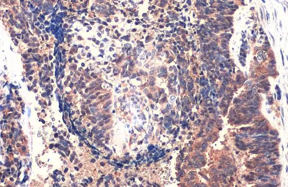
MMP1 antibody detects MMP1 protein at cytoplasm by immunohistochemical analysis. Sample: Paraffin-embedded human endometrial carcinoma. MMP1 stained by MMP1 antibody (GTX100534) diluted at 1:500. Antigen Retrieval: Citrate buffer, pH 6.0, 15 min
 diluted at 1:500. Antigen Retrieval: Citrate buffer, pH 6.0, 15 min")
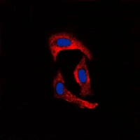
![MMP1 antibody detects MMP1 protein at cytoplasm by immunofluorescent analysis. Sample: HUVEC cells were fixed in 4% paraformaldehyde at RT for 15 min. Green: MMP1 stained by MMP1 antibody (GTX100534) diluted at 1:500. Red: alpha Tubulin, a cytoskeleton marker, stained by alpha Tubulin antibody [GT114] (GTX628802) diluted at 1:1000. Blue: Fluoroshield with DAPI (GTX30920).](https://www.genetex.com/upload/website/prouct_img/normal/GTX100534/GTX100534_44321_20220513_ICC_IF_w_23060100_599.webp "MMP1 antibody detects MMP1 protein at cytoplasm by immunofluorescent analysis. Sample: HUVEC cells were fixed in 4% paraformaldehyde at RT for 15 min. Green: MMP1 stained by MMP1 antibody (GTX100534) diluted at 1:500. Red: alpha Tubulin, a cytoskeleton marker, stained by alpha Tubulin antibody [GT114] (GTX628802) diluted at 1:1000. Blue: Fluoroshield with DAPI (GTX30920).")
 diluted at 1:500. Antigen Retrieval: Citrate buffer, pH 6.0, 15 min")


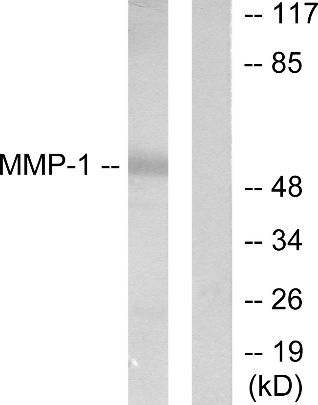
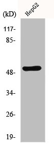
 were separated by 10% SDS-PAGE, and the membrane was blotted with MMP1 antibody (GTX100534) diluted at 1:1000. The HRP-conjugated anti-rabbit IgG antibody (GTX213110-01) was used to detect the primary antibody. Corresponding RNA expression data are based on Human Protein Atlas program.")
MMP1 antibody detects MMP1 protein at cytoplasm by immunohistochemical analysis. Sample: Paraffin-embedded human endometrial carcinoma. MMP1 stained by MMP1 antibody (GTX100534) diluted at 1:500. Antigen Retrieval: Citrate buffer, pH 6.0, 15 min
MMP1 antibody
GTX100534
ApplicationsImmunoFluorescence, Western Blot, ImmunoCytoChemistry, ImmunoHistoChemistry, ImmunoHistoChemistry Paraffin
Product group Antibodies
ReactivityHuman, Mouse
TargetMMP1
Overview
- SupplierGeneTex
- Product NameMMP1 antibody
- Delivery Days Customer9
- Application Supplier NoteWB: 1:500-1:3000. IHC-P: 1:100-1:1000. *Optimal dilutions/concentrations should be determined by the researcher.Not tested in other applications.
- ApplicationsImmunoFluorescence, Western Blot, ImmunoCytoChemistry, ImmunoHistoChemistry, ImmunoHistoChemistry Paraffin
- CertificationResearch Use Only
- ClonalityPolyclonal
- Concentration1 mg/ml
- ConjugateUnconjugated
- Gene ID4312
- Target nameMMP1
- Target descriptionmatrix metallopeptidase 1
- Target synonymsCLG, CLGN, interstitial collagenase, fibroblast collagenase, matrix metallopeptidase 1 (interstitial collagenase), matrix metalloprotease 1
- HostRabbit
- IsotypeIgG
- Protein IDP03956
- Protein NameInterstitial collagenase
- Scientific DescriptionProteins of the matrix metalloproteinase (MMP) family are involved in the breakdown of extracellular matrix in normal physiological processes, such as embryonic development, reproduction, and tissue remodeling, as well as in disease processes, such as arthritis and metastasis. Most MMPs are secreted as inactive proproteins which are activated when cleaved by extracellular proteinases. This gene encodes a secreted enzyme which breaks down the interstitial collagens, types I, II, and III. The gene is part of a cluster of MMP genes which localize to chromosome 11q22.3. Alternative splicing results in multiple transcript variants.[provided by RefSeq]
- ReactivityHuman, Mouse
- Storage Instruction-20°C or -80°C,2°C to 8°C
- UNSPSC41116161
Datasheet
Related products
Product group Antibodies
Anti-MMP-1 AntibodyA100981
ApplicationsWestern Blot, ELISA
ReactivityHuman
- SizePrice

Product group Antibodies
Anti-MMP1 Antibody Picoband(r)A00733-1-CARRIER-FREE
ApplicationsFlow Cytometry, ImmunoFluorescence, Western Blot, ELISA, ImmunoCytoChemistry, ImmunoHistoChemistry
ReactivityHuman
TargetMMP1
- SizePrice
Product group Antibodies

MMP1 Antibody (Preservative Free)LS-C743708
ApplicationsELISA
ReactivityHuman
TargetMMP1
- SizePrice
Product group Antibodies
MMP1 AntibodyCSB-PA003252
ApplicationsImmunoFluorescence, Western Blot, ELISA, ImmunoHistoChemistry
ReactivityHuman
TargetMMP1
- SizePrice
Product group Antibodies
MMP1 Polyclonal AntibodyCAC14096
ApplicationsWestern Blot, ELISA, ImmunoHistoChemistry
TargetMMP1
- SizePrice

Product group Antibodies
References
MMP-1 Polyclonal Antibodybs-4597R
ApplicationsImmunoFluorescence, Western Blot, ELISA, ImmunoCytoChemistry, ImmunoHistoChemistry, ImmunoHistoChemistry Frozen, ImmunoHistoChemistry Paraffin
ReactivityBovine, Canine, Equine, Human, Mouse, Rabbit, Rat, Sheep
TargetMMP1
- SizePrice
Product group Antibodies
MMP1 antibody [F15 P3 B6]GTX15771
ApplicationsWestern Blot
ReactivityHuman
TargetMMP1
- SizePrice
Product group Antibodies
References
MMP1 antibodyGTX00674
ApplicationsImmunoFluorescence, Western Blot, ImmunoCytoChemistry, ImmunoHistoChemistry, ImmunoHistoChemistry Paraffin
ReactivityHuman, Mouse, Rat
TargetMMP1
- SizePrice