

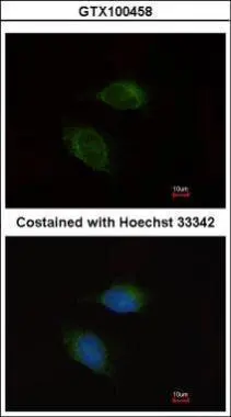
Immunofluorescence analysis of methanol-fixed HeLa, using MMP9(GTX100458) antibody at 1:200 dilution.
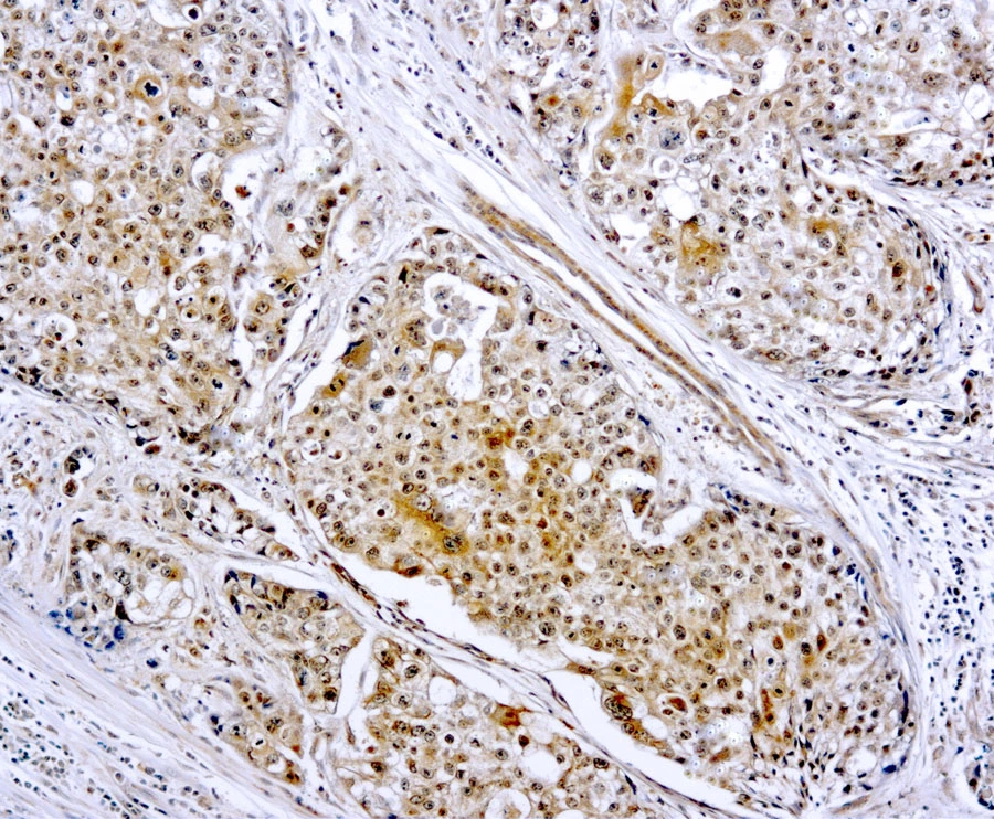
![MMP9 antibody [N2C1], Internal detects MMP9 protein at cytoplasm by immunohistochemical analysis. Sample: Paraffin-embedded mouse lymph node. MMP9 stained by MMP9 antibody [N2C1], Internal (GTX100458) diluted at 1:500. Antigen Retrieval: Citrate buffer, pH 6.0, 15 min](https://www.genetex.com/upload/website/prouct_img/normal/GTX100458/GTX100458_43138_20190719_IHC-P_M_w_23060100_920.webp "MMP9 antibody [N2C1], Internal detects MMP9 protein at cytoplasm by immunohistochemical analysis. Sample: Paraffin-embedded mouse lymph node. MMP9 stained by MMP9 antibody [N2C1], Internal (GTX100458) diluted at 1:500. Antigen Retrieval: Citrate buffer, pH 6.0, 15 min")
![Untreated (–) and treated (+) MCF-7 whole cell extracts (30 μg) were separated by 7.5% SDS-PAGE, and the membrane was blotted with MMP9 antibody [N2C1], Internal (GTX100458) diluted at 1:1000. The HRP-conjugated anti-rabbit IgG antibody (GTX213110-01) was used to detect the primary antibody.](https://www.genetex.com/upload/website/prouct_img/normal/GTX100458/GTX100458_45301_20240322_WB_treatment_PMA_Brefeldin_24032600_289.webp "Untreated (–) and treated (+) MCF-7 whole cell extracts (30 μg) were separated by 7.5% SDS-PAGE, and the membrane was blotted with MMP9 antibody [N2C1], Internal (GTX100458) diluted at 1:1000. The HRP-conjugated anti-rabbit IgG antibody (GTX213110-01) was used to detect the primary antibody.")
 in 2020.")


![Untreated (–) and treated (+) U2OS whole cell extracts (30 μg) were separated by 7.5% SDS-PAGE, and the membrane was blotted with MMP9 antibody [N2C1], Internal (GTX100458) diluted at 1:1000. The HRP-conjugated anti-rabbit IgG antibody (GTX213110-01) was used to detect the primary antibody.](https://www.genetex.com/upload/website/prouct_img/normal/GTX100458/GTX100458_45301_20240524_WB_treatment_TPA_24052802_216.webp "Untreated (–) and treated (+) U2OS whole cell extracts (30 μg) were separated by 7.5% SDS-PAGE, and the membrane was blotted with MMP9 antibody [N2C1], Internal (GTX100458) diluted at 1:1000. The HRP-conjugated anti-rabbit IgG antibody (GTX213110-01) was used to detect the primary antibody.")
![Mouse tissue extract (50 μg) was separated by 7.5% SDS-PAGE, and the membrane was blotted with MMP9 antibody [N2C1], Internal (GTX100458) diluted at 1:1000. The HRP-conjugated anti-rabbit IgG antibody (GTX213110-01) was used to detect the primary antibody.](https://www.genetex.com/upload/website/prouct_img/normal/GTX100458/GTX100458_45301_20240607_WB_M_spleen_24061301_801.webp "Mouse tissue extract (50 μg) was separated by 7.5% SDS-PAGE, and the membrane was blotted with MMP9 antibody [N2C1], Internal (GTX100458) diluted at 1:1000. The HRP-conjugated anti-rabbit IgG antibody (GTX213110-01) was used to detect the primary antibody.")
![U87-MG whole cell extract and conditioned medium (30 μg) were separated by 7.5% SDS-PAGE, and the membrane was blotted with MMP9 antibody [N2C1], Internal (GTX100458) diluted at 1:1000. The HRP-conjugated anti-rabbit IgG antibody (GTX213110-01) was used to detect the primary antibody.](https://www.genetex.com/upload/website/prouct_img/normal/GTX100458/GTX100458_45476_20240719_WB_Fraction_25061003_784.webp "U87-MG whole cell extract and conditioned medium (30 μg) were separated by 7.5% SDS-PAGE, and the membrane was blotted with MMP9 antibody [N2C1], Internal (GTX100458) diluted at 1:1000. The HRP-conjugated anti-rabbit IgG antibody (GTX213110-01) was used to detect the primary antibody.")
Immunofluorescence analysis of methanol-fixed HeLa, using MMP9(GTX100458) antibody at 1:200 dilution.
MMP9 antibody [N2C1], Internal
GTX100458
ApplicationsImmunoFluorescence, Western Blot, ImmunoCytoChemistry, ImmunoHistoChemistry, ImmunoHistoChemistry Frozen, ImmunoHistoChemistry Paraffin
Product group Antibodies
ReactivityBovine, Human, Mouse, Rat
TargetMMP9
Overview
- SupplierGeneTex
- Product NameMMP9 antibody [N2C1], Internal
- Delivery Days Customer9
- Application Supplier NoteWB: 1:500-1:3000. ICC/IF: 1:100-1:1000. IHC-P: 1:100-1:1000. *Optimal dilutions/concentrations should be determined by the researcher.Not tested in other applications.
- ApplicationsImmunoFluorescence, Western Blot, ImmunoCytoChemistry, ImmunoHistoChemistry, ImmunoHistoChemistry Frozen, ImmunoHistoChemistry Paraffin
- CertificationResearch Use Only
- ClonalityPolyclonal
- Concentration1.96 mg/ml
- ConjugateUnconjugated
- Gene ID4318
- Target nameMMP9
- Target descriptionmatrix metallopeptidase 9
- Target synonymsCLG4B, GELB, MANDP2, MMP-9, matrix metalloproteinase-9, macrophage gelatinase, matrix metallopeptidase 9 (gelatinase B, 92kDa gelatinase, 92kDa type IV collagenase), matrix metalloproteinase 9 (gelatinase B, 92kDa gelatinase, 92kDa type IV collagenase), type V collagenase
- HostRabbit
- IsotypeIgG
- Protein IDP14780
- Protein NameMatrix metalloproteinase-9
- Scientific DescriptionProteins of the matrix metalloproteinase (MMP) family are involved in the breakdown of extracellular matrix in normal physiological processes, such as embryonic development, reproduction, and tissue remodeling, as well as in disease processes, such as arthritis and metastasis. Most MMPs are secreted as inactive proproteins which are activated when cleaved by extracellular proteinases. The enzyme encoded by this gene degrades type IV and V collagens. Studies in rhesus monkeys suggest that the enzyme is involved in IL-8-induced mobilization of hematopoietic progenitor cells from bone marrow, and murine studies suggest a role in tumor-associated tissue remodeling. [provided by RefSeq]
- ReactivityBovine, Human, Mouse, Rat
- Storage Instruction-20°C or -80°C,2°C to 8°C
- UNSPSC41116161
Datasheet
Related products
Product group Antibodies
Anti-MMP9 AntibodyA101376
ApplicationsWestern Blot, ELISA
ReactivityHuman
- SizePrice
Product group Antibodies
Anti-MMP9 Antibody130-10537
ApplicationsELISA
ReactivityHuman
TargetMMP9
- SizePrice
Product group Antibodies

Anti-gelatinase B [REGA-3G12]AB04197-1.1
ApplicationsImmunoPrecipitation, Western Blot, ELISA, Neutralisation/Blocking
ReactivityHuman
TargetMMP9
- SizePrice

Product group Antibodies
Anti-MMP9 AntibodyAMAB90804
ApplicationsWestern Blot, ImmunoHistoChemistry
ReactivityHuman
TargetMMP9
- SizePrice
Product group Antibodies

MMP9 / Gelatinase B AntibodyLS-C831545
ApplicationsImmunoHistoChemistry
ReactivityHuman, Mouse
TargetMMP9
- SizePrice
Product group Antibodies
MMP9 AntibodyCSB-PA002676
ApplicationsImmunoFluorescence, Western Blot, ELISA, ImmunoHistoChemistry
ReactivityHuman
TargetMMP9
- SizePrice
Product group Antibodies
ApplicationsFlow Cytometry
TargetMMP9
- SizePrice

Product group Antibodies
References
MMP9 Polyclonal AntibodyBS-4593R
ApplicationsFlow Cytometry, ImmunoFluorescence, Western Blot, ELISA, ImmunoCytoChemistry, ImmunoHistoChemistry, ImmunoHistoChemistry Frozen, ImmunoHistoChemistry Paraffin
ReactivityHuman
TargetMMP9
- SizePrice
Product group Antibodies
MMP9 antibodyGTX22167
ApplicationsImmunoHistoChemistry, ImmunoHistoChemistry Paraffin
ReactivityHuman
TargetMMP9
- SizePrice