

Mouse anti Lamin B1
MUB1103P-CE/IVD
ApplicationsFlow Cytometry, Western Blot, ELISA, ImmunoCytoChemistry, ImmunoHistoChemistry, ImmunoHistoChemistry Frozen
Product group Antibodies
ReactivityBovine, Canine, Human, Mouse, Rabbit, Rat, Sheep, Zebra Fish
TargetLMNB1
Overview
- SupplierNordic-MUbio
- Product NameMouse anti Lamin B1
- Delivery Days Customer7
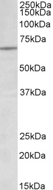
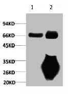
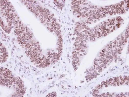
- Application Supplier Note119D5-F1 is suitable for immunocytochemistry on permeabilised cells, immunohistochemistry on frozen tissues, immunoblotting, ELISA and flow cytometry. Optimal antibody dilution should be determined by titration; recommended range is 1:100 - 1:200 for flow cytometry, immunocytochemistry and for immunohistochemistry with avidin-biotinylated Horseradish peroxidase complex (ABC) as detection reagent, and 1:100 - 1:1000 for immunoblotting applications.
- ApplicationsFlow Cytometry, Western Blot, ELISA, ImmunoCytoChemistry, ImmunoHistoChemistry, ImmunoHistoChemistry Frozen
- Applications SupplierFlow Cytometry;Immunocytochemistry;Western Blotting;Immunohistochemistry (frozen);ELISA
- CertificationCE-IVD
- ClonalityMonoclonal
- Clone ID119D5-F1
- Gene ID4001
- Target nameLMNB1
- Target descriptionlamin B1
- Target synonymsADLD, ADLDAT, LMN, LMN2, LMNB, MCPH26, lamin-B1
- HostMouse
- IsotypeIgG1
- Protein IDP20700
- Protein NameLamin-B1
- Source119D5-F1 is a Mouse monoclonal IgG1/k antibody derived by fusion of P3/X63.Ag8.653 Mouse myeloma cells with spleen cells from a BALB/c Mouse immunized with purified Rat liver lamins.
- ReactivityBovine, Canine, Human, Mouse, Rabbit, Rat, Sheep, Zebra Fish
- Reactivity SupplierBovine;Canine;Human;Mouse;Rabbit;Rat;Sheep;Zebrafish
- UNSPSC12352203
Related products
Product group Antibodies
Anti-Lamin B1 AntibodyA82688
ApplicationsWestern Blot, ELISA
ReactivityHuman
- SizePrice
Product group Antibodies
Anti-LMNB1 Antibody144-01910
ApplicationsImmunoFluorescence, Western Blot, ImmunoHistoChemistry
ReactivityHuman, Mouse, Rat
TargetLMNB1
- SizePrice

Product group Antibodies
Anti-LMNB1 AntibodyAMAB91251
ApplicationsWestern Blot, ImmunoCytoChemistry, ImmunoHistoChemistry
ReactivityHuman
TargetLMNB1
- SizePrice
Product group Antibodies

LMNB1 / Lamin B1 Antibody (clone 4K2)LS-C767832
ApplicationsImmunoFluorescence, ImmunoPrecipitation, Western Blot, ImmunoHistoChemistry, ImmunoHistoChemistry Paraffin
ReactivityHuman, Mouse, Rat
TargetLMNB1
- SizePrice
Product group Antibodies
References
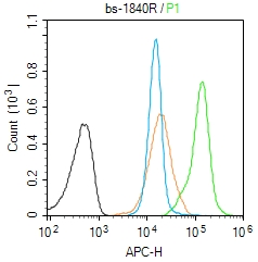
Lamin B1 Polyclonal AntibodyBS-1840R
ApplicationsFlow Cytometry, ImmunoFluorescence, Western Blot, ELISA, ImmunoCytoChemistry, ImmunoHistoChemistry, ImmunoHistoChemistry Frozen, ImmunoHistoChemistry Paraffin
ReactivityMouse
TargetLMNB1
- SizePrice
Product group Antibodies
LMNB1 Monoclonal AntibodyCSB-MA000191
ApplicationsImmunoPrecipitation, Western Blot, ELISA
ReactivityHuman, Mouse, Rat
TargetLMNB1
- SizePrice
Product group Antibodies

ApplicationsWestern Blot, ELISA
ReactivityBovine, Human, Porcine
TargetLMNB1
- SizePrice
Product group Antibodies
ApplicationsImmunoPrecipitation, Western Blot, ImmunoCytoChemistry, ImmunoHistoChemistry
ReactivityBovine, Canine, Equine, Goat, Mouse, Porcine, Rat
TargetLMNB1
- SizePrice
Product group Antibodies
Lamin B1 antibodyGTX103292
ApplicationsImmunoPrecipitation, Western Blot, ImmunoHistoChemistry, ImmunoHistoChemistry Paraffin
ReactivityHamster, Human, Mouse, Rat
TargetLMNB1
- SizePrice