Mouse anti Vimentin
MUB1902P-CE/IVD
ApplicationsFlow Cytometry, Western Blot, ImmunoCytoChemistry, ImmunoHistoChemistry, ImmunoHistoChemistry Frozen, ImmunoHistoChemistry Paraffin
Product group Antibodies
ReactivityChicken, Human, Mammals, Rat, Sheep
TargetVIM
Overview
- SupplierNordic-MUbio
- Product NameMouse anti Vimentin
- Delivery Days Customer7
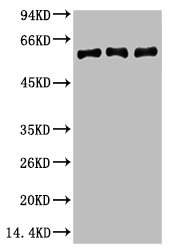
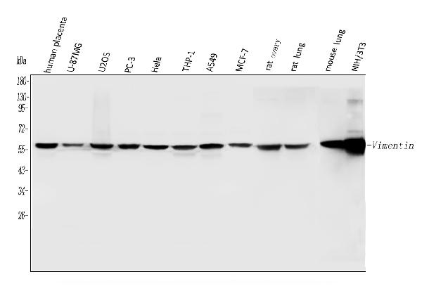
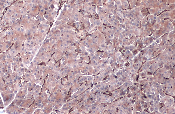
- Application Supplier NoteV9 is suitable for immunoblotting, immunocytochemistry, immunohistochemistry on frozen and paraffin-embedded tissues and flow cytometry. For staining paraffin-embedded tissues pretreatment by boiling tissue sections for 10 minutues in 10 mM citRate buffer (pH 6.0) is required. Optimal antibody dilution should be determined by titration; recommended range is 1:25 - 1:200 for flow cytometry, and for immunohistochemistry with avidin-biotinylated Horseradish peroxidase complex (ABC) as detection reagent, and 1:100 - 1:1000 for immunoblotting applications.
- ApplicationsFlow Cytometry, Western Blot, ImmunoCytoChemistry, ImmunoHistoChemistry, ImmunoHistoChemistry Frozen, ImmunoHistoChemistry Paraffin
- Applications SupplierFlow Cytometry;Immunocytochemistry;Immunohistochemistry (frozen);Immunohistochemistry (paraffin);Western Blotting
- CertificationCE-IVD
- ClonalityMonoclonal
- Clone IDV9
- Gene ID7431
- Target nameVIM
- Target descriptionvimentin
- Target synonymsvimentin, epididymis secretory sperm binding protein
- HostMouse
- IsotypeIgG1
- Protein IDP08670
- Protein NameVimentin
- SourceV9 is a Mouse monoclonal IgG1 antibody derived by fusion of PAI Mouse myeloma cells with spleen cells from a BALB/c Mouse immunized with vimentin isolated from porcine lens.
- ReactivityChicken, Human, Mammals, Rat, Sheep
- Reactivity SupplierChicken;Human;Rat;Sheep
- UNSPSC12352203


