

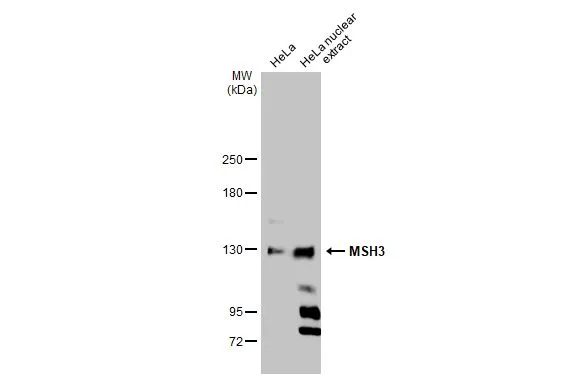
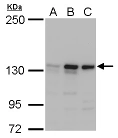
HeLa whole cell and nuclear extracts (30 μg) were separated by 5% SDS-PAGE, and the membrane was blotted with MSH3 antibody [C2C3], C-term (GTX113781) diluted at 1:1000. The HRP-conjugated anti-rabbit IgG antibody (GTX213110-01) was used to detect the primary antibody.
![MSH3 antibody [C2C3], C-term detects MSH3 protein at nucleus by immunohistochemical analysis. Sample: Paraffin-embedded human lung cancer. MSH3 stained by MSH3 antibody [C2C3], C-term (GTX113781) diluted at 1:600. Antigen Retrieval: Citrate buffer, pH 6.0, 15 min](https://www.genetex.com/upload/website/prouct_img/normal/GTX113781/GTX113781_43635_20190719_IHC-P_w_23060501_138.webp "MSH3 antibody [C2C3], C-term detects MSH3 protein at nucleus by immunohistochemical analysis. Sample: Paraffin-embedded human lung cancer. MSH3 stained by MSH3 antibody [C2C3], C-term (GTX113781) diluted at 1:600. Antigen Retrieval: Citrate buffer, pH 6.0, 15 min")
 antibody at 1:500 dilution.
Antigen Retrieval: Trilogy? (EDTA based, pH 8.0) buffer, 15min")
![MSH3 antibody [C2C3], C-term detects MSH3 protein at nucleus by immunofluorescent analysis. Sample: HeLa cells were fixed in 4% paraformaldehyde at RT for 15 min. Green: MSH3 stained by MSH3 antibody [C2C3], C-term (GTX113781) diluted at 1:500. Blue: Fluoroshield with DAPI (GTX30920). Scale bar= 10 μm.](https://www.genetex.com/upload/website/prouct_img/normal/GTX113781/GTX113781_43635_20200311_ICC_IF_w_23060501_253.webp "MSH3 antibody [C2C3], C-term detects MSH3 protein at nucleus by immunofluorescent analysis. Sample: HeLa cells were fixed in 4% paraformaldehyde at RT for 15 min. Green: MSH3 stained by MSH3 antibody [C2C3], C-term (GTX113781) diluted at 1:500. Blue: Fluoroshield with DAPI (GTX30920). Scale bar= 10 μm.")
![A549 whole cell and nuclear extracts (30 μg) were separated by 5% SDS-PAGE, and the membrane was blotted with MSH3 antibody [C2C3], C-term (GTX113781) diluted at 1:1000. The HRP-conjugated anti-rabbit IgG antibody (GTX213110-01) was used to detect the primary antibody.](https://www.genetex.com/upload/website/prouct_img/normal/GTX113781/GTX113781_43775_20231027_WB_Fraction_23110819_462.webp "A549 whole cell and nuclear extracts (30 μg) were separated by 5% SDS-PAGE, and the membrane was blotted with MSH3 antibody [C2C3], C-term (GTX113781) diluted at 1:1000. The HRP-conjugated anti-rabbit IgG antibody (GTX213110-01) was used to detect the primary antibody.")
HeLa whole cell and nuclear extracts (30 μg) were separated by 5% SDS-PAGE, and the membrane was blotted with MSH3 antibody [C2C3], C-term (GTX113781) diluted at 1:1000. The HRP-conjugated anti-rabbit IgG antibody (GTX213110-01) was used to detect the primary antibody.
MSH3 antibody [C2C3], C-term
GTX113781
ApplicationsImmunoFluorescence, Western Blot, ImmunoCytoChemistry, ImmunoHistoChemistry, ImmunoHistoChemistry Paraffin
Product group Antibodies
ReactivityHuman
TargetMSH3
Overview
- SupplierGeneTex
- Product NameMSH3 antibody [C2C3], C-term
- Delivery Days Customer9
- Application Supplier NoteWB: 1:500-1:3000. ICC/IF: 1:100-1:1000. IHC-P: 1:100-1:1000. *Optimal dilutions/concentrations should be determined by the researcher.Not tested in other applications.
- ApplicationsImmunoFluorescence, Western Blot, ImmunoCytoChemistry, ImmunoHistoChemistry, ImmunoHistoChemistry Paraffin
- CertificationResearch Use Only
- ClonalityPolyclonal
- Concentration1.79 mg/ml
- ConjugateUnconjugated
- Gene ID4437
- Target nameMSH3
- Target descriptionmutS homolog 3
- Target synonymsDUP, FAP4, MRP1, DNA mismatch repair protein Msh3, divergent upstream protein, epididymis secretory sperm binding protein, hMSH3, mismatch repair protein 1
- HostRabbit
- IsotypeIgG
- Protein IDP20585
- Protein NameDNA mismatch repair protein Msh3
- Scientific DescriptionComponent of the post-replicative DNA mismatch repair system (MMR). Heterodimerizes with MSH2 to form MutS beta which binds to DNA mismatches thereby initiating DNA repair. When bound, the MutS beta heterodimer bends the DNA helix and shields approximately 20 base pairs. MutS beta recognizes large insertion-deletion loops (IDL) up to 13 nucleotides long. After mismatch binding, forms a ternary complex with the MutL alpha heterodimer, which is thought to be responsible for directing the downstream MMR events, including strand discrimination, excision, and resynthesis.
- ReactivityHuman
- Storage Instruction-20°C or -80°C,2°C to 8°C
- UNSPSC41116161
Datasheet
Related products
Product group Antibodies
Anti-MSH3 (A80) AntibodyA26260
ApplicationsWestern Blot, ImmunoHistoChemistry
ReactivityHuman, Mouse, Rat
- SizePrice

Product group Antibodies
Anti-MSH3 (A80) AntibodyA02455
ApplicationsWestern Blot, ImmunoHistoChemistry
ReactivityHuman, Mouse, Rat
TargetMSH3
- SizePrice

Product group Antibodies
Anti-MSH3 AntibodyAMAB91910
ApplicationsWestern Blot, ImmunoCytoChemistry, ImmunoHistoChemistry
ReactivityHuman
TargetMSH3
- SizePrice
Product group Antibodies
MSH3 Polyclonal AntibodyBS-4919R
ApplicationsImmunoFluorescence, Western Blot, ImmunoHistoChemistry, ImmunoHistoChemistry Frozen, ImmunoHistoChemistry Paraffin
ReactivityBovine, Equine, Human, Mouse, Porcine, Rabbit, Rat
TargetMSH3
- SizePrice
Product group Antibodies
MSH3 AntibodyCSB-PA003320
ApplicationsWestern Blot, ELISA, ImmunoHistoChemistry
ReactivityHuman
TargetMSH3
- SizePrice
Product group Antibodies
MSH3 antibody [N1N2], N-termGTX103228
ApplicationsWestern Blot
ReactivityHuman
TargetMSH3
- SizePrice
Product group Antibodies

MSH3 AntibodyLS-C334210
ApplicationsWestern Blot, ImmunoHistoChemistry
ReactivityHuman
TargetMSH3
- SizePrice
![Non-transfected (–) and transfected (+) 293T whole cell extracts were separated by 5% SDS-PAGE, and the membrane was blotted with MSH3 antibody [HL2546] (GTX638918) diluted at 1:4000. The HRP-conjugated anti-rabbit IgG antibody (GTX213110-01) was used to detect the primary antibody.](https://www.genetex.com/upload/website/prouct_img/normal/GTX638918/GTX638918_T-45131_20231013_WB_B_23101921_744.webp)
Product group Antibodies
MSH3 antibody [HL2546]GTX638918
ApplicationsWestern Blot
ReactivityHuman
TargetMSH3
- SizePrice
Product group Antibodies
MSH3 antibodyGTX32728
ApplicationsWestern Blot
ReactivityHuman
TargetMSH3
- SizePrice
Product group Antibodies
anti-MSH3 (human), Rabbit Monoclonal (RM405)REV-31-1291-00
ApplicationsWestern Blot, ImmunoHistoChemistry
ReactivityHuman
TargetMSH3
- SizePrice