

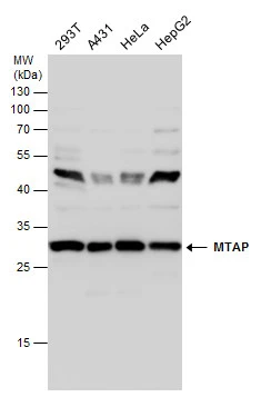
MTAP antibody detects MTAP protein by Western blot analysis. Various whole cell extracts (30 μg) were separated by 12% SDS-PAGE, and the membrane was blotted with MTAP antibody (GTX109117) diluted at a dilution of 1:5000.
 antibody at 1:500 dilution.
Antigen Retrieval: Trilogy? (EDTA based, pH 8.0) buffer, 15min")
![MTAP antibody [N1C3] detects MTAP protein at cytoplasm and nucleus by immunofluorescent analysis. Sample: HeLa cells were fixed in 4% paraformaldehyde at RT for 15 min. Green: MTAP protein stained by MTAP antibody [N1C3] (GTX109117) diluted at 1:500. Blue: Hoechst 33342 staining.](https://www.genetex.com/upload/website/prouct_img/normal/GTX109117/GTX109117_42032_20151030_IFA_w_23060120_539.webp "MTAP antibody [N1C3] detects MTAP protein at cytoplasm and nucleus by immunofluorescent analysis. Sample: HeLa cells were fixed in 4% paraformaldehyde at RT for 15 min. Green: MTAP protein stained by MTAP antibody [N1C3] (GTX109117) diluted at 1:500. Blue: Hoechst 33342 staining.")
![Immunoprecipitation of MTAP protein from HeLa whole cell extracts using 5 μg of MTAP antibody [N1C3] (GTX109117). Western blot analysis was performed using MTAP antibody [N1C3] (GTX109117). EasyBlot anti-Rabbit IgG (GTX221666-01) was used as a secondary reagent.](https://www.genetex.com/upload/website/prouct_img/normal/GTX109117/GTX109117_40023_20150529_IP_w_23060120_795.webp "Immunoprecipitation of MTAP protein from HeLa whole cell extracts using 5 μg of MTAP antibody [N1C3] (GTX109117). Western blot analysis was performed using MTAP antibody [N1C3] (GTX109117). EasyBlot anti-Rabbit IgG (GTX221666-01) was used as a secondary reagent.")
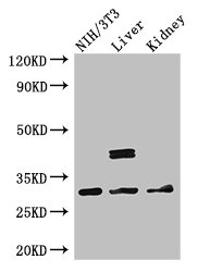
 A: mouse liver 12% SDS PAGE GTX109117 diluted at 1:1000 The HRP-conjugated anti-rabbit IgG antibody (GTX213110-01) was used to detect the primary antibody.")
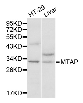
MTAP antibody detects MTAP protein by Western blot analysis. Various whole cell extracts (30 μg) were separated by 12% SDS-PAGE, and the membrane was blotted with MTAP antibody (GTX109117) diluted at a dilution of 1:5000.
MTAP antibody [N1C3]
GTX109117
ApplicationsImmunoFluorescence, ImmunoPrecipitation, Western Blot, ImmunoCytoChemistry, ImmunoHistoChemistry, ImmunoHistoChemistry Paraffin
Product group Antibodies
ReactivityHuman, Mouse
TargetMTAP
Overview
- SupplierGeneTex
- Product NameMTAP antibody [N1C3]
- Delivery Days Customer9
- Application Supplier NoteWB: 1:500-1:3000. ICC/IF: 1:100-1:1000. IHC-P: 1:100-1:1000. IP: 1:100-1:500. *Optimal dilutions/concentrations should be determined by the researcher.Not tested in other applications.
- ApplicationsImmunoFluorescence, ImmunoPrecipitation, Western Blot, ImmunoCytoChemistry, ImmunoHistoChemistry, ImmunoHistoChemistry Paraffin
- CertificationResearch Use Only
- ClonalityPolyclonal
- Concentration0.18 mg/ml
- ConjugateUnconjugated
- Gene ID4507
- Target nameMTAP
- Target descriptionmethylthioadenosine phosphorylase
- Target synonymsBDMF, DMSFH, DMSMFH, HEL-249, LGMBF, MSAP, c86fus, S-methyl-5'-thioadenosine phosphorylase, 5'-methylthioadenosine phosphorylase, MTA phosphorylase, MTAPase, MeSAdo phosphorylase, epididymis luminal protein 249, epididymis secretory sperm binding protein
- HostRabbit
- IsotypeIgG
- Protein IDQ13126
- Protein NameS-methyl-5'-thioadenosine phosphorylase
- Scientific DescriptionThis gene encodes an enzyme that plays a major role in polyamine metabolism and is important for the salvage of both adenine and methionine. The encoded enzyme is deficient in many cancers because this gene and the tumor suppressor p16 gene are co-deleted. Multiple alternatively spliced transcript variants have been described for this gene, but their full-length natures remain unknown. [provided by RefSeq]
- ReactivityHuman, Mouse
- Storage Instruction-20°C or -80°C,2°C to 8°C
- UNSPSC41116161
Datasheet
Related products

Product group Antibodies
Anti-MTAP Antibody Picoband(r)A05448-2-CARRIER-FREE
ApplicationsFlow Cytometry, ImmunoFluorescence, Western Blot, ELISA, ImmunoCytoChemistry
ReactivityHuman, Mouse, Rat
TargetMTAP
- SizePrice
Product group Antibodies
Anti-MTAP AntibodyA30601
ApplicationsImmunoFluorescence, Western Blot, ImmunoHistoChemistry
ReactivityHuman, Mouse, Rat
- SizePrice
Product group Antibodies
Anti-MTAP Antibody144-01049
ApplicationsImmunoFluorescence, Western Blot, ImmunoHistoChemistry
ReactivityHuman, Mouse, Rat
TargetMTAP
- SizePrice
Product group Antibodies

MTAP AntibodyLS-C832068
ApplicationsELISA, ImmunoHistoChemistry
ReactivityHuman
TargetMTAP
- SizePrice
Product group Antibodies
ApplicationsFlow Cytometry, Western Blot, ImmunoCytoChemistry
ReactivityHuman, Mouse, Rat
TargetMTAP
- SizePrice
Product group Antibodies
MTAP AntibodyCSB-PA622639LA01HU
ApplicationsImmunoFluorescence, Western Blot, ELISA
ReactivityHuman, Mouse
TargetMTAP
- SizePrice
Product group Antibodies
MTAP Polyclonal AntibodyCAC14661
ApplicationsImmunoFluorescence, Western Blot, ELISA
ReactivityMouse
TargetMTAP
- SizePrice
Product group Antibodies
MTAP antibody [MTAP/1813]GTX17975
ApplicationsWestern Blot, ELISA, ImmunoHistoChemistry, ImmunoHistoChemistry Paraffin, Other Application
ReactivityHuman
TargetMTAP
- SizePrice
![WB analysis of various samples using GTX02677 MTAP antibody [MTAP/3137R]. Lane 1 : HeLa whole cell lysate Lane 2 : A431 whole cell lysate Lane 3 : HepG2 whole cell lysate Lane 4 : HAP1 whole cell lysate Lane 5 : U937 whole cell lysate](https://www.genetex.com/upload/website/prouct_img/normal/GTX02677/GTX02677_20210319_WB_w_23053122_480.webp)
Product group Antibodies
MTAP antibody [MTAP/3137R]GTX02677
ApplicationsWestern Blot, ELISA, ImmunoHistoChemistry, ImmunoHistoChemistry Paraffin
ReactivityHuman
TargetMTAP
- SizePrice
![IHC-P analysis of human non-invasive papillary urothelial carcinoma (NIPUC) tissue using GTX04964 MTAP antibody [MSVA-741R] HistoMAX?. Non invasive urothelial carcinoma pTa with complete absence of MTAP staining in all tumor cells. MTAP positive stroma cells serve as an internal control.](https://www.genetex.com/upload/website/prouct_img/normal/GTX04964/GTX04964_20241028_IHC-P_24102820_319.webp)
Product group Antibodies
ApplicationsImmunoHistoChemistry, ImmunoHistoChemistry Paraffin
ReactivityHuman
TargetMTAP
- SizePrice