

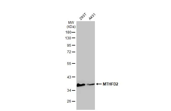
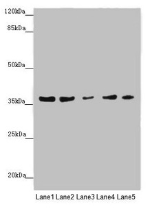
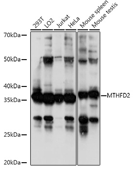
Various whole cell extracts (30 μg) were separated by 10% SDS-PAGE, and the membrane was blotted with MTHFD2 antibody [N1C3] (GTX104990) diluted at 1:1000. The HRP-conjugated anti-rabbit IgG antibody (GTX213110-01) was used to detect the primary antibody.
![MTHFD2 antibody [N1C3] detects MTHFD2 protein at mitochondria by immunohistochemical analysis. Sample: Paraffin-embedded human MCF7 xenograft. MTHFD2 antibody [N1C3] (GTX104990) diluted at 1:500.
Antigen Retrieval: Citrate buffer, pH 6.0, 15 min](https://www.genetex.com/upload/website/prouct_img/normal/GTX104990/GTX104990_40093_20151227_IHC-P_w_23060120_452.webp "MTHFD2 antibody [N1C3] detects MTHFD2 protein at mitochondria by immunohistochemical analysis. Sample: Paraffin-embedded human MCF7 xenograft. MTHFD2 antibody [N1C3] (GTX104990) diluted at 1:500.
Antigen Retrieval: Citrate buffer, pH 6.0, 15 min")
![Immunoprecipitation of MTHFD2 protein from 293T whole cell extracts using 5 μg of MTHFD2 antibody [N1C3] (GTX104990). Western blot analysis was performed using MTHFD2 antibody [N1C3] (GTX104990). EasyBlot anti-Rabbit IgG (GTX221666-01) was used as a secondary reagent.](https://www.genetex.com/upload/website/prouct_img/normal/GTX104990/GTX104990_40093_20150420_IP_w_23060120_542.webp "Immunoprecipitation of MTHFD2 protein from 293T whole cell extracts using 5 μg of MTHFD2 antibody [N1C3] (GTX104990). Western blot analysis was performed using MTHFD2 antibody [N1C3] (GTX104990). EasyBlot anti-Rabbit IgG (GTX221666-01) was used as a secondary reagent.")
 antibody at 1:500 dilution.
Antigen Retrieval: Trilogy? (EDTA based, pH 8.0) buffer, 15min")
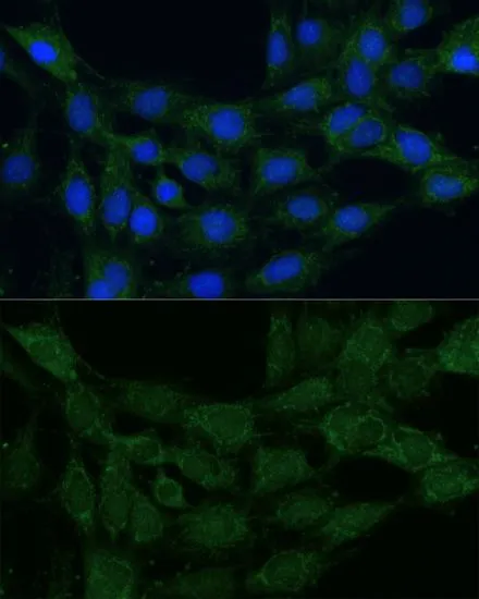
![MTHFD2 antibody [N1C3] detects MTHFD2 protein at mitochondria by immunofluorescent analysis. Sample: HeLa cells were fixed in 4% paraformaldehyde at RT for 15 min. Green: MTHFD2 stained by MTHFD2 antibody [N1C3] (GTX104990) diluted at 1:500. Blue: Fluoroshield with DAPI (GTX30920).](https://www.genetex.com/upload/website/prouct_img/normal/GTX104990/GTX104990_43670_20200429_ICC_IF_w_23060120_531.webp "MTHFD2 antibody [N1C3] detects MTHFD2 protein at mitochondria by immunofluorescent analysis. Sample: HeLa cells were fixed in 4% paraformaldehyde at RT for 15 min. Green: MTHFD2 stained by MTHFD2 antibody [N1C3] (GTX104990) diluted at 1:500. Blue: Fluoroshield with DAPI (GTX30920).")
![Non-transfected (–) and transfected (+) 293T whole cell extracts (30 μg) were separated by 10% SDS-PAGE, and the membrane was blotted with MTHFD2 antibody [N1C3] (GTX104990) diluted at 1:3000. The HRP-conjugated anti-rabbit IgG antibody (GTX213110-01) was used to detect the primary antibody.](https://www.genetex.com/upload/website/prouct_img/normal/GTX104990/GTX104990_40093_20181228_WB_shRNA_watermark_w_23060120_713.webp "Non-transfected (–) and transfected (+) 293T whole cell extracts (30 μg) were separated by 10% SDS-PAGE, and the membrane was blotted with MTHFD2 antibody [N1C3] (GTX104990) diluted at 1:3000. The HRP-conjugated anti-rabbit IgG antibody (GTX213110-01) was used to detect the primary antibody.")
![MTHFD2 antibody [N1C3] detects MTHFD2 protein at mitochondria in human breast carcinoma by immunohistochemical analysis. Sample: Paraffin-embedded human breast carcinoma. MTHFD2 antibody [N1C3] (GTX104990) diluted at 1:500.
Antigen Retrieval: Citrate buffer, pH 6.0, 15 min](https://www.genetex.com/upload/website/prouct_img/normal/GTX104990/GTX104990_40093_20151223_IHC-P_w_23060120_842.webp "MTHFD2 antibody [N1C3] detects MTHFD2 protein at mitochondria in human breast carcinoma by immunohistochemical analysis. Sample: Paraffin-embedded human breast carcinoma. MTHFD2 antibody [N1C3] (GTX104990) diluted at 1:500.
Antigen Retrieval: Citrate buffer, pH 6.0, 15 min")
![MTHFD2 antibody [N1C3] detects MTHFD2 protein at mitochondria by immunohistochemical analysis. Sample: Paraffin-embedded human colon cancer. MTHFD2 stained by MTHFD2 antibody [N1C3] (GTX104990) diluted at 1:500. Antigen Retrieval: Citrate buffer, pH 6.0, 15 min](https://www.genetex.com/upload/website/prouct_img/normal/GTX104990/GTX104990_43593_20210226_IHC-P_w_23060120_872.webp "MTHFD2 antibody [N1C3] detects MTHFD2 protein at mitochondria by immunohistochemical analysis. Sample: Paraffin-embedded human colon cancer. MTHFD2 stained by MTHFD2 antibody [N1C3] (GTX104990) diluted at 1:500. Antigen Retrieval: Citrate buffer, pH 6.0, 15 min")
Various whole cell extracts (30 μg) were separated by 10% SDS-PAGE, and the membrane was blotted with MTHFD2 antibody [N1C3] (GTX104990) diluted at 1:1000. The HRP-conjugated anti-rabbit IgG antibody (GTX213110-01) was used to detect the primary antibody.
MTHFD2 antibody [N1C3]
GTX104990
ApplicationsImmunoFluorescence, ImmunoPrecipitation, Western Blot, ImmunoCytoChemistry, ImmunoHistoChemistry, ImmunoHistoChemistry Paraffin
Product group Antibodies
ReactivityHuman
TargetMTHFD2
Overview
- SupplierGeneTex
- Product NameMTHFD2 antibody [N1C3]
- Delivery Days Customer9
- Application Supplier NoteWB: 1:500-1:3000. ICC/IF: 1:100-1:1000. IHC-P: 1:100-1:1000. IP: 1:100-1:500. *Optimal dilutions/concentrations should be determined by the researcher.Not tested in other applications.
- ApplicationsImmunoFluorescence, ImmunoPrecipitation, Western Blot, ImmunoCytoChemistry, ImmunoHistoChemistry, ImmunoHistoChemistry Paraffin
- CertificationResearch Use Only
- ClonalityPolyclonal
- Concentration0.13 mg/ml
- ConjugateUnconjugated
- Gene ID10797
- Target nameMTHFD2
- Target descriptionmethylenetetrahydrofolate dehydrogenase (NADP+ dependent) 2, methenyltetrahydrofolate cyclohydrolase
- Target synonymsNMDMC, bifunctional methylenetetrahydrofolate dehydrogenase/cyclohydrolase, mitochondrial, NAD-dependent methylene tetrahydrofolate dehydrogenase cyclohydrolase
- HostRabbit
- IsotypeIgG
- Protein IDP13995
- Protein NameBifunctional methylenetetrahydrofolate dehydrogenase/cyclohydrolase, mitochondrial
- Scientific DescriptionThis gene encodes a nuclear-encoded mitochondrial bifunctional enzyme with methylenetetrahydrofolate dehydrogenase and methenyltetrahydrofolate cyclohydrolase activities. The enzyme functions as a homodimer and is unique in its absolute requirement for magnesium and inorganic phosphate. Formation of the enzyme-magnesium complex allows binding of NAD. Alternative splicing results in two different transcripts, one protein-coding and the other not protein-coding. This gene has a pseudogene on chromosome 7. [provided by RefSeq]
- ReactivityHuman
- Storage Instruction-20°C or -80°C,2°C to 8°C
- UNSPSC41116161
Datasheet
Related products
Product group Antibodies
MTHFD2 AntibodyCSB-PA015156ESR1HU
ApplicationsWestern Blot, ELISA, ImmunoHistoChemistry
ReactivityHuman
TargetMTHFD2
- SizePrice
Product group Antibodies
Anti-MTHFD2 AntibodyA11302
ApplicationsImmunoFluorescence, Western Blot, ImmunoCytoChemistry, ImmunoHistoChemistry
ReactivityHuman, Mouse, Rat
- SizePrice
Product group Antibodies

MTHFD2 AntibodyLS-C830994
ApplicationsELISA, ImmunoHistoChemistry
ReactivityHuman, Mouse
TargetMTHFD2
- SizePrice

Product group Antibodies
Anti-MTHFD2 AntibodyHPA049657
ApplicationsImmunoHistoChemistry
ReactivityHuman
TargetMTHFD2
- SizePrice
Product group Antibodies
MTHFD2 antibodyGTX64519
ApplicationsImmunoFluorescence, Western Blot, ImmunoCytoChemistry, ImmunoHistoChemistry, ImmunoHistoChemistry Paraffin
ReactivityHuman, Mouse, Rat
TargetMTHFD2
- SizePrice
Product group Antibodies
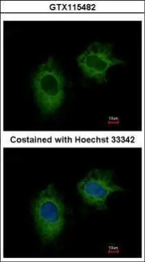
MTHFD2 antibody [N3C3]GTX115482
ApplicationsImmunoFluorescence, Western Blot, ImmunoCytoChemistry, ImmunoHistoChemistry, ImmunoHistoChemistry Paraffin
ReactivityHuman, Mouse, Rat
TargetMTHFD2
- SizePrice
Product group Antibodies
Anti-MTHFD2 Antibody144-10386
ApplicationsWestern Blot
ReactivityHuman, Mouse
TargetMTHFD2
- SizePrice