

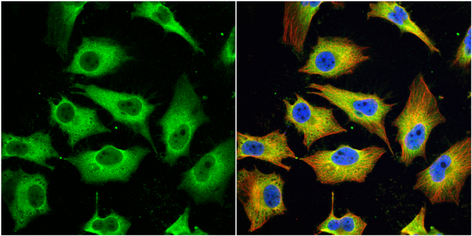
MX1 antibody [N2C2], Internal detects MX1 protein at cytoplasm by immunofluorescent analysis. Sample: HeLa cells were fixed in 4% paraformaldehyde at RT for 15 min. Green: MX1 protein stained by MX1 antibody [N2C2], Internal (GTX110256) diluted at 1:200. Red: alpha Tubulin, a cytoskeleton marker, stained by alpha Tubulin antibody [GT114] (GTX628802) diluted at 1:1000. Blue: Hoechst 33342 staining.
![MX1 antibody [N2C2], Internal detects MX1 protein at cytoplasm in human colon carcinoma by immunohistochemical analysis. Sample: Paraffin-embedded human colon carcinoma. MX1 antibody [N2C2], Internal (GTX110256) diluted at 1:500.
Antigen Retrieval: Citrate buffer, pH 6.0, 15 min](https://www.genetex.com/upload/website/prouct_img/normal/GTX110256/GTX110256_40073_20160125_IHC-P_w_23060500_210.webp "MX1 antibody [N2C2], Internal detects MX1 protein at cytoplasm in human colon carcinoma by immunohistochemical analysis. Sample: Paraffin-embedded human colon carcinoma. MX1 antibody [N2C2], Internal (GTX110256) diluted at 1:500.
Antigen Retrieval: Citrate buffer, pH 6.0, 15 min")
![MX1 antibody [N2C2], Internal detects MX1 protein by western blot analysis. A. 30 μg 293T whole cell lysate/extract B. 30 μg whole cell lysate/extract of human MX1-transfected 293T cells. 7.5% SDS-PAGE MX1 antibody [N2C2], Internal (GTX110256) dilution: 1:10000 The HRP-conjugated anti-rabbit IgG antibody (GTX213110-01) was used to detect the primary antibody.](https://www.genetex.com/upload/website/prouct_img/normal/GTX110256/GTX110256_40037_WB_B_w_23060500_172.webp "MX1 antibody [N2C2], Internal detects MX1 protein by western blot analysis. A. 30 μg 293T whole cell lysate/extract B. 30 μg whole cell lysate/extract of human MX1-transfected 293T cells. 7.5% SDS-PAGE MX1 antibody [N2C2], Internal (GTX110256) dilution: 1:10000 The HRP-conjugated anti-rabbit IgG antibody (GTX213110-01) was used to detect the primary antibody.")
 antibody at 1:500 dilution.
Antigen Retrieval: Citrate buffer, pH 6.0, 15 min")


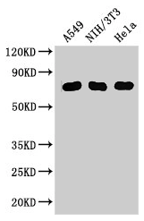
![Various whole cell extracts (30 μg) were separated by 7.5% SDS-PAGE, and the membrane was blotted with MX1 antibody [N2C2], Internal (GTX110256) diluted at 1:1000. The HRP-conjugated anti-rabbit IgG antibody (GTX213110-01) was used to detect the primary antibody. Corresponding RNA expression data for the same cell lines are based on Human Protein Atlas program.](https://www.genetex.com/upload/website/prouct_img/normal/GTX110256/GTX110256_45637_20241227_WB_TPM_watermark_25041720_758.webp "Various whole cell extracts (30 μg) were separated by 7.5% SDS-PAGE, and the membrane was blotted with MX1 antibody [N2C2], Internal (GTX110256) diluted at 1:1000. The HRP-conjugated anti-rabbit IgG antibody (GTX213110-01) was used to detect the primary antibody. Corresponding RNA expression data for the same cell lines are based on Human Protein Atlas program.")
MX1 antibody [N2C2], Internal detects MX1 protein at cytoplasm by immunofluorescent analysis. Sample: HeLa cells were fixed in 4% paraformaldehyde at RT for 15 min. Green: MX1 protein stained by MX1 antibody [N2C2], Internal (GTX110256) diluted at 1:200. Red: alpha Tubulin, a cytoskeleton marker, stained by alpha Tubulin antibody [GT114] (GTX628802) diluted at 1:1000. Blue: Hoechst 33342 staining.
MX1 antibody [N2C2], Internal
GTX110256
ApplicationsImmunoFluorescence, Western Blot, ImmunoCytoChemistry, ImmunoHistoChemistry, ImmunoHistoChemistry Frozen, ImmunoHistoChemistry Paraffin
Product group Antibodies
ReactivityBovine, Human, Monkey, Rabbit
TargetMX1
Overview
- SupplierGeneTex
- Product NameMX1 antibody [N2C2], Internal
- Delivery Days Customer9
- Application Supplier NoteWB: 1:500-1:3000. ICC/IF: 1:100-1:1000. IHC-P: 1:100-1:1000. *Optimal dilutions/concentrations should be determined by the researcher.Not tested in other applications.
- ApplicationsImmunoFluorescence, Western Blot, ImmunoCytoChemistry, ImmunoHistoChemistry, ImmunoHistoChemistry Frozen, ImmunoHistoChemistry Paraffin
- CertificationResearch Use Only
- ClonalityPolyclonal
- Concentration1.23 mg/ml
- ConjugateUnconjugated
- Gene ID4599
- Target nameMX1
- Target descriptionMX dynamin like GTPase 1
- Target synonymsIFI-78K, IFI78, MX, MxA, lncMX1-215, interferon-induced GTP-binding protein Mx1, interferon-induced protein p78, interferon-inducible protein p78, interferon-regulated resistance GTP-binding protein MxA, myxoma resistance protein 1, myxovirus (influenza virus) resistance 1, interferon-inducible protein p78
- HostRabbit
- IsotypeIgG
- Protein IDP20591
- Protein NameInterferon-induced GTP-binding protein Mx1
- Scientific DescriptionIn mouse, the interferon-inducible Mx protein is responsible for a specific antiviral state against influenza virus infection. The protein encoded by this gene is similar to the mouse protein as determined by its antigenic relatedness, induction conditions, physicochemical properties, and amino acid analysis. This cytoplasmic protein is a member of both the dynamin family and the family of large GTPases. Two transcript variants encoding the same protein have been found for this gene. [provided by RefSeq]
- ReactivityBovine, Human, Monkey, Rabbit
- Storage Instruction-20°C or -80°C,2°C to 8°C
- UNSPSC41116161
Datasheet
Related products
Product group Antibodies
MX1 AntibodyCSB-PA015249LA01HU
ApplicationsImmunoFluorescence, ImmunoPrecipitation, Western Blot, ELISA, ImmunoHistoChemistry
ReactivityHuman, Mouse
TargetMX1
- SizePrice

Product group Antibodies
Anti-MX1 Antibody Picoband(r)A00849-1-CARRIER-FREE
ApplicationsFlow Cytometry, Western Blot, ELISA
ReactivityHuman, Mouse
TargetMX1
- SizePrice
Product group Antibodies
Anti-MX1 AntibodyA11481
ApplicationsImmunoFluorescence, Western Blot, ImmunoCytoChemistry, ImmunoHistoChemistry
ReactivityHuman, Mouse
- SizePrice
Product group Antibodies
Anti-MX1 Antibody144-65170
ApplicationsWestern Blot
ReactivityHuman, Mouse
TargetMX1
- SizePrice

Product group Antibodies
Anti-MX1 AntibodyHPA030917
ApplicationsWestern Blot, ImmunoHistoChemistry
ReactivityHuman
TargetMX1
- SizePrice
Product group Antibodies

MX1 / MX AntibodyLS-C331685
ApplicationsWestern Blot
ReactivityHuman, Mouse
TargetMX1
- SizePrice
Product group Antibodies
MX1 Monoclonal AntibodyBSM-51528M
ApplicationsWestern Blot
ReactivityHuman, Porcine
TargetMX1
- SizePrice
![Non-transfected (–) and transfected (+) 293T whole cell extracts (30 μg) were separated by 7.5% SDS-PAGE, and the membrane was blotted with MX1 antibody [HL1790] (GTX637442) diluted at 1:5000. The HRP-conjugated anti-rabbit IgG antibody (GTX213110-01) was used to detect the primary antibody.](https://www.genetex.com/upload/website/prouct_img/normal/GTX637442/GTX637442_T-44809_20221223_WB_multiple_B_22122722_968.webp)
Product group Antibodies
MX1 antibody [HL1790]GTX637442
ApplicationsImmunoFluorescence, Western Blot, ImmunoCytoChemistry, ImmunoHistoChemistry, ImmunoHistoChemistry Paraffin
ReactivityFeline, Human
TargetMX1
- SizePrice
![MX1 antibody [HL2051] detects MX1 protein at cytoplasm and nucleus by immunohistochemical analysis. Sample: Paraffin-embedded human lung cancer. MX1 stained by MX1 antibody [HL2051] (GTX637955) diluted at 1:100. Antigen Retrieval: Citrate buffer, pH 6.0, 15 min](https://www.genetex.com/upload/website/prouct_img/normal/GTX637955/GTX637955_T-44886_20221230_IHC-P_23020621_426.webp)
Product group Antibodies
MX1 antibody [HL2051]GTX637955
ApplicationsImmunoFluorescence, Western Blot, ImmunoCytoChemistry, ImmunoHistoChemistry, ImmunoHistoChemistry Paraffin
ReactivityCanine, Feline, Human, Mouse
TargetMX1
- SizePrice