

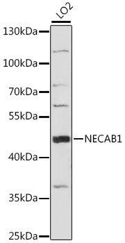
NECAB1 Antibody (aa208-236)
LS-C164816
ApplicationsWestern Blot
Product group Antibodies
ReactivityHuman
TargetNECAB1
Overview
- SupplierLifeSpan BioSciences
- Product NameNECAB1 Antibody (aa208-236)
- Delivery Days Customer23
- ApplicationsWestern Blot
- Applications SupplierWB (1:1000)
- CertificationResearch Use Only
- ClonalityPolyclonal
- ConjugateUnconjugated
- Estimated Purity...
- Gene ID64168
- Target nameNECAB1
- Target descriptionN-terminal EF-hand calcium binding protein 1
- Target synonymsEFCBP1, STIP-1, N-terminal EF-hand calcium-binding protein 1, EF-hand calcium-binding protein 1, neuronal calcium binding protein, neuronal calcium-binding protein 1, synaptotagmin interacting protein 1
- HostRabbit
- ReactivityHuman
- Storage Instruction-20°C,2°C to 8°C
- UNSPSC41116161
Related products
Product group Antibodies
Anti-NECAB1 Antibody144-63957
ApplicationsWestern Blot
ReactivityHuman, Mouse, Rat
TargetNECAB1
- SizePrice

Product group Antibodies
Anti-NECAB1 AntibodyAMAB90798
ApplicationsWestern Blot, ImmunoHistoChemistry
ReactivityHuman
TargetNECAB1
- SizePrice

Product group Antibodies
Anti-NECAB1 Antibody Picoband(r)A13973-1-CARRIER-FREE
ApplicationsImmunoFluorescence, Western Blot, ELISA, ImmunoCytoChemistry, ImmunoHistoChemistry
ReactivityHuman, Mouse, Rat
TargetNECAB1
- SizePrice
Product group Antibodies
Anti-NECAB1 AntibodyA89965
ApplicationsWestern Blot
ReactivityHuman, Mouse, Rat
- SizePrice
Product group Antibodies
NECAB1 AntibodyCSB-PA843298LA01HU
ApplicationsImmunoFluorescence, ELISA
ReactivityHuman
TargetNECAB1
- SizePrice