

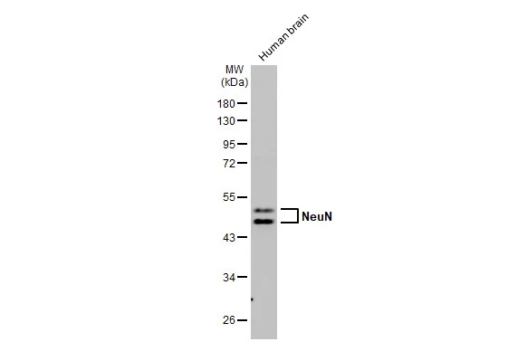
Human tissue extract (30 μg) was separated by 10% SDS-PAGE, and the membrane was blotted with NeuN antibody [HL2550] (GTX638922) diluted at 1:1000. The HRP-conjugated anti-rabbit IgG antibody (GTX213110-01) was used to detect the primary antibody, and the signal was developed with Trident ECL plus-Enhanced.
![Various tissue extracts (50 μg) were separated by 10% SDS-PAGE, and the membrane was blotted with NeuN antibody [HL2550] (GTX638922) diluted at 1:1000. The HRP-conjugated anti-rabbit IgG antibody (GTX213110-01) was used to detect the primary antibody, and the signal was developed with Trident ECL plus-Enhanced.](https://www.genetex.com/upload/website/prouct_img/normal/GTX638922/GTX638922_T-45131_20230825_WB_R_tissue_23083020_976.webp "Various tissue extracts (50 μg) were separated by 10% SDS-PAGE, and the membrane was blotted with NeuN antibody [HL2550] (GTX638922) diluted at 1:1000. The HRP-conjugated anti-rabbit IgG antibody (GTX213110-01) was used to detect the primary antibody, and the signal was developed with Trident ECL plus-Enhanced.")
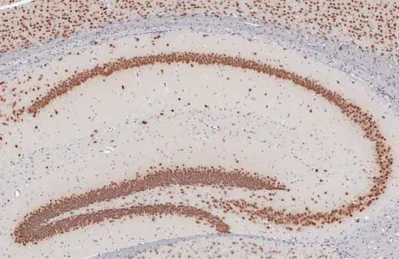
![NeuN antibody [HL2550] detects NeuN protein by immunohistochemical analysis. Sample: Paraffin-embedded rat tissues. NeuN stained by NeuN antibody [HL2550] (GTX638922) diluted at 1:100. Antigen Retrieval: Citrate buffer, pH 6.0, 15 min](https://www.genetex.com/upload/website/prouct_img/normal/GTX638922/GTX638922_T-45131_20230829_IHC-P_multiple_R_23091319_382.webp "NeuN antibody [HL2550] detects NeuN protein by immunohistochemical analysis. Sample: Paraffin-embedded rat tissues. NeuN stained by NeuN antibody [HL2550] (GTX638922) diluted at 1:100. Antigen Retrieval: Citrate buffer, pH 6.0, 15 min")
![NeuN antibody [HL2550] detects NeuN protein by immunohistochemical analysis. Sample: Paraffin-embedded mouse tissues. NeuN stained by NeuN antibody [HL2550] (GTX638922) diluted at 1:100. Antigen Retrieval: Citrate buffer, pH 6.0, 15 min](https://www.genetex.com/upload/website/prouct_img/normal/GTX638922/GTX638922_T-45131_20230829_IHC-P_multiple_M_23091319_704.webp "NeuN antibody [HL2550] detects NeuN protein by immunohistochemical analysis. Sample: Paraffin-embedded mouse tissues. NeuN stained by NeuN antibody [HL2550] (GTX638922) diluted at 1:100. Antigen Retrieval: Citrate buffer, pH 6.0, 15 min")
![Various tissue extracts (50 μg) were separated by 10% SDS-PAGE, and the membrane was blotted with NeuN antibody [HL2550] (GTX638922) diluted at 1:2000. The HRP-conjugated anti-rabbit IgG antibody (GTX213110-01) was used to detect the primary antibody, and the signal was developed with Trident ECL plus-Enhanced.](https://www.genetex.com/upload/website/prouct_img/normal/GTX638922/GTX638922_45201_20231027_WB_M_tissue_23103019_959.webp "Various tissue extracts (50 μg) were separated by 10% SDS-PAGE, and the membrane was blotted with NeuN antibody [HL2550] (GTX638922) diluted at 1:2000. The HRP-conjugated anti-rabbit IgG antibody (GTX213110-01) was used to detect the primary antibody, and the signal was developed with Trident ECL plus-Enhanced.")
![NeuN antibody [HL2550] detects NeuN protein at cytoplasm and nucleus by immunohistochemical analysis. Sample: Frozen-sectioned mouse cerebellum. Green: NeuN stained by NeuN antibody [HL2550] (GTX638922) diluted at 1:100. Antigen Retrieval: 4% paraformaldehyde](https://www.genetex.com/upload/website/prouct_img/normal/GTX638922/GTX638922_T-45131_20231222_IHC-Fr_M_24021917_897.webp "NeuN antibody [HL2550] detects NeuN protein at cytoplasm and nucleus by immunohistochemical analysis. Sample: Frozen-sectioned mouse cerebellum. Green: NeuN stained by NeuN antibody [HL2550] (GTX638922) diluted at 1:100. Antigen Retrieval: 4% paraformaldehyde")
Human tissue extract (30 μg) was separated by 10% SDS-PAGE, and the membrane was blotted with NeuN antibody [HL2550] (GTX638922) diluted at 1:1000. The HRP-conjugated anti-rabbit IgG antibody (GTX213110-01) was used to detect the primary antibody, and the signal was developed with Trident ECL plus-Enhanced.
NeuN antibody [HL2550]
GTX638922
ApplicationsWestern Blot, ImmunoHistoChemistry, ImmunoHistoChemistry Frozen, ImmunoHistoChemistry Paraffin
Product group Antibodies
ReactivityHuman, Mouse, Rat
TargetRbfox3
Overview
- SupplierGeneTex
- Product NameNeuN antibody [HL2550]
- Delivery Days Customer9
- Application Supplier NoteWB: 1:500-1:3000. *Optimal dilutions/concentrations should be determined by the researcher.Not tested in other applications.
- ApplicationsWestern Blot, ImmunoHistoChemistry, ImmunoHistoChemistry Frozen, ImmunoHistoChemistry Paraffin
- CertificationResearch Use Only
- ClonalityMonoclonal
- Clone IDHL2550
- Concentration1 mg/ml
- ConjugateUnconjugated
- Gene ID52897
- Target nameRbfox3
- Target descriptionRNA binding protein, fox-1 homolog (C. elegans) 3
- Target synonymsFox-3, Hrnbp3, NeuN, Neuna60, RNA binding protein fox-1 homolog 3, fox-1 homolog C, hexaribonucleotide binding protein 3, neuN antigen, neuronal nuclear antigen A60, neuronal nuclei antigen
- HostRabbit
- IsotypeIgG
- Protein IDQ8BIF2
- Protein NameRNA binding protein fox-1 homolog 3
- ReactivityHuman, Mouse, Rat
- Storage Instruction-20°C or -80°C,2°C to 8°C
- UNSPSC41116161
Datasheet
Related products

Product group Antibodies
Anti-NeuN/Rbfox3 Antibody Picoband(r)A11954-1-CARRIER-FREE
ApplicationsImmunoFluorescence, Western Blot, ELISA, ImmunoHistoChemistry
ReactivityMouse, Rat
TargetRbfox3
- SizePrice
Product group Antibodies
Rbfox3 AntibodyCSB-PA806847ZA01MO
ApplicationsWestern Blot, ELISA
ReactivityMouse
TargetRbfox3
- SizePrice
![IHC-P analysis of rat cerebellum tissue using GTX30773 NeuN antibody [2Q158].Immunoreactivity is seen as nuclear staining in the neurons in the granular layer. Antigen retireval : Citrate Buffer, pH 6.0 Dilution : 1:100](https://www.genetex.com/upload/website/prouct_img/normal/GTX30773/GTX30773_20191203_IHC-P_29_w_23060722_654.webp)
Product group Antibodies
References
NeuN antibody [2Q158]GTX30773
ApplicationsImmunoFluorescence, Western Blot, ImmunoCytoChemistry, ImmunoHistoChemistry, ImmunoHistoChemistry Frozen, ImmunoHistoChemistry Paraffin
ReactivityCanine, Human, Mouse, Rabbit, Reptile, Rat
TargetRbfox3
- SizePrice
Product group Antibodies
NeuN antibody [A60]GTX01767
ApplicationsFlow Cytometry, ImmunoFluorescence, Western Blot, ImmunoCytoChemistry, ImmunoHistoChemistry, ImmunoHistoChemistry Frozen, ImmunoHistoChemistry Paraffin
ReactivityHuman, Mouse, Rabbit, Rat
TargetRbfox3
- SizePrice
Product group Antibodies
NeuN antibodyGTX133127
ApplicationsImmunoFluorescence, ImmunoCytoChemistry, ImmunoHistoChemistry, ImmunoHistoChemistry Frozen, ImmunoHistoChemistry Paraffin
ReactivityMouse, Rat
TargetRbfox3
- SizePrice