

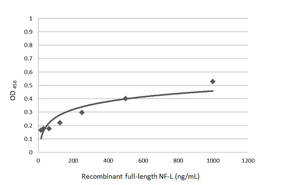
Sandwich ELISA detection of recombinant full-length NF-L protein using GTX101142 as capture antibody at concentration of 5 μg/mL and GTX60544 as detection antibody was diluted at 1:10000. Mouse IgG antibody (HRP) (GTX213111-01) was diluted at 1:10000 and used to detect the primary antibody.
 antibody at 1:500 dilution.
Antigen Retrieval: Trilogy? (EDTA based, pH 8.0) buffer, 15min")
![NF-L antibody detects NF-L protein at cytoplasm by immunofluorescent analysis. Sample: DIV9 rat E18 primary cortical neurons were fixed in 4% paraformaldehyde at RT for 15 min. Green: NF-L protein stained by NF-L antibody (GTX101142) diluted at 1:500. Red: beta Tubulin 3/ Tuj1, stained by beta Tubulin 3/ Tuj1 antibody [GT11710] (GTX631836) diluted at 1:500. Blue: Fluoroshield with DAPI (GTX30920).](https://www.genetex.com/upload/website/prouct_img/normal/GTX101142/GTX101142_40737_20170503_IFA_R_w_23060100_702.webp "NF-L antibody detects NF-L protein at cytoplasm by immunofluorescent analysis. Sample: DIV9 rat E18 primary cortical neurons were fixed in 4% paraformaldehyde at RT for 15 min. Green: NF-L protein stained by NF-L antibody (GTX101142) diluted at 1:500. Red: beta Tubulin 3/ Tuj1, stained by beta Tubulin 3/ Tuj1 antibody [GT11710] (GTX631836) diluted at 1:500. Blue: Fluoroshield with DAPI (GTX30920).")
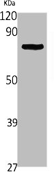
 was separated by 7.5% SDS-PAGE, and the membrane was blotted with NF-L antibody (GTX101142) diluted at 1:500. The HRP-conjugated anti-rabbit IgG antibody (GTX213110-01) was used to detect the primary antibody.")
 (GTX213111-01) was diluted at 1:10000 and used to detect the primary antibody.")
 was separated by 7.5% SDS-PAGE, and the membrane was blotted with NF-L antibody (GTX101142) diluted at 1:1000. The HRP-conjugated anti-rabbit IgG antibody (GTX213110-01) was used to detect the primary antibody.")
![NF-L antibody detects NF-L protein expression by immunohistochemical analysis. Sample: Frozen-sectioned adult mouse cerebellum. Green: NF-L protein stained by NF-L antibody (GTX101142) diluted at 1:250. Red: beta Tubulin 3/ TUJ1, stained by beta Tubulin 3/ TUJ1 antibody [GT11710] (GTX631836) diluted at 1:500. Blue: Fluoroshield with DAPI (GTX30920).](https://www.genetex.com/upload/website/prouct_img/normal/GTX101142/GTX101142_40737_20170531_IHC-Fr_M_w_23060100_238.webp "NF-L antibody detects NF-L protein expression by immunohistochemical analysis. Sample: Frozen-sectioned adult mouse cerebellum. Green: NF-L protein stained by NF-L antibody (GTX101142) diluted at 1:250. Red: beta Tubulin 3/ TUJ1, stained by beta Tubulin 3/ TUJ1 antibody [GT11710] (GTX631836) diluted at 1:500. Blue: Fluoroshield with DAPI (GTX30920).")
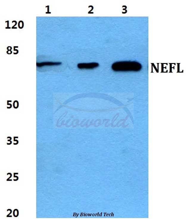
 were separated by 7.5% SDS-PAGE, and the membrane was blotted with NF-L antibody (GTX101142) diluted at 1:1000. The HRP-conjugated anti-rabbit IgG antibody (GTX213110-01) was used to detect the primary antibody. Corresponding RNA expression data for the same cell lines are based on Human Protein Atlas program.")
 diluted at 1:1000. The HRP-conjugated anti-rabbit IgG antibody (GTX213110-01) was used to detect the primary antibody.")
![NF-L antibody detects NF-L protein by immunofluorescent analysis. Sample: Neuro2A cells were fixed in 4% paraformaldehyde at RT for 15 min. Green: NF-L stained by NF-L antibody (GTX101142) diluted at 1:500. Red: alpha Tubulin, a cytoskeleton marker, stained by alpha Tubulin antibody [GT114] (GTX628802) diluted at 1:1000. Blue: Fluoroshield with DAPI (GTX30920).](https://www.genetex.com/upload/website/prouct_img/normal/GTX101142/GTX101142_45113_20241018_ICC_IF_M_24110700_125.webp "NF-L antibody detects NF-L protein by immunofluorescent analysis. Sample: Neuro2A cells were fixed in 4% paraformaldehyde at RT for 15 min. Green: NF-L stained by NF-L antibody (GTX101142) diluted at 1:500. Red: alpha Tubulin, a cytoskeleton marker, stained by alpha Tubulin antibody [GT114] (GTX628802) diluted at 1:1000. Blue: Fluoroshield with DAPI (GTX30920).")
Sandwich ELISA detection of recombinant full-length NF-L protein using GTX101142 as capture antibody at concentration of 5 μg/mL and GTX60544 as detection antibody was diluted at 1:10000. Mouse IgG antibody (HRP) (GTX213111-01) was diluted at 1:10000 and used to detect the primary antibody.
NF-L antibody
GTX101142
ApplicationsImmunoFluorescence, Western Blot, ELISA, ImmunoCytoChemistry, ImmunoHistoChemistry, ImmunoHistoChemistry Frozen, ImmunoHistoChemistry Paraffin
Product group Antibodies
ReactivityHuman, Mouse, Rat
TargetNEFL
Overview
- SupplierGeneTex
- Product NameNF-L antibody
- Delivery Days Customer9
- Application Supplier NoteWB: 1:500-1:20000. ICC/IF: 1:100-1:1000. IHC-P: 1:100-1:1000. IHC-Fr: 1:100-1:1000. *Optimal dilutions/concentrations should be determined by the researcher.Not tested in other applications.
- ApplicationsImmunoFluorescence, Western Blot, ELISA, ImmunoCytoChemistry, ImmunoHistoChemistry, ImmunoHistoChemistry Frozen, ImmunoHistoChemistry Paraffin
- CertificationResearch Use Only
- ClonalityPolyclonal
- Concentration1.09 mg/ml
- ConjugateUnconjugated
- Gene ID4747
- Target nameNEFL
- Target descriptionneurofilament light chain
- Target synonymsCMT1F, CMT2E, CMTDIG, NF-L, NF68, NFL, PPP1R110, neurofilament light polypeptide, light molecular weight neurofilament protein, neurofilament protein, light chain, neurofilament subunit NF-L, neurofilament triplet L protein, neurofilament, light polypeptide 68kDa, protein phosphatase 1, regulatory subunit 110
- HostRabbit
- IsotypeIgG
- Protein IDP07196
- Protein NameNeurofilament light polypeptide
- Scientific DescriptionNeurofilaments are type IV intermediate filament heteropolymers composed of light, medium, and heavy chains. Neurofilaments comprise the axoskeleton and they functionally maintain the neuronal caliber. They may also play a role in intracellular transport to axons and dendrites. This gene encodes the light chain neurofilament protein. Mutations in this gene cause Charcot-Marie-Tooth disease types 1F (CMT1F) and 2E (CMT2E), disorders of the peripheral nervous system that are characterized by distinct neuropathies. A pseudogene has been identified on chromosome Y. [provided by RefSeq]
- ReactivityHuman, Mouse, Rat
- Storage Instruction-20°C or -80°C,2°C to 8°C
- UNSPSC41116161
Datasheet
Related products
Product group Antibodies
NEFL AntibodyCSB-PA004743
ApplicationsWestern Blot, ELISA
ReactivityHuman, Mouse, Rat
TargetNEFL
- SizePrice

Product group Antibodies
Anti-NEFL AntibodyAMAB91314
ApplicationsWestern Blot, ImmunoCytoChemistry, ImmunoHistoChemistry
ReactivityHuman, Mouse, Rat
TargetNEFL
- SizePrice
Product group Antibodies

Anti-Neurofilament light [MS-C2]AB00946-10.0-BT
ApplicationsELISA
ReactivityHuman
TargetNEFL
- SizePrice
Product group Antibodies
Anti-NEFL AntibodyA28063
ApplicationsWestern Blot
ReactivityHuman, Mouse, Rat
- SizePrice

Product group Antibodies
Anti-NEFL/NF-L Antibody Picoband(r)A02482-1-CARRIER-FREE
ApplicationsFlow Cytometry, ImmunoFluorescence, Western Blot, ELISA, ImmunoHistoChemistry
ReactivityHuman, Mouse, Rat
TargetNEFL
- SizePrice
Product group Antibodies

Neurofilament AntibodyLS-C835096
ApplicationsImmunoHistoChemistry
ReactivityHuman
- SizePrice
Product group Antibodies

Goat anti-NEFL (aa330-343)EB13051
ApplicationsWestern Blot, ELISA
ReactivityBovine, Canine, Human, Mouse, Porcine, Rat
TargetNEFL
- SizePrice
Product group Antibodies

Mouse anti Neurofilament 70 kDMUB1303P
ApplicationsWestern Blot, ImmunoHistoChemistry, ImmunoHistoChemistry Frozen, ImmunoHistoChemistry Paraffin
ReactivityHuman
TargetNEFL
- SizePrice
Product group Antibodies
Nefl Polyclonal AntibodyCAC08666
ApplicationsImmunoFluorescence, ImmunoPrecipitation, ELISA, ImmunoHistoChemistry
TargetNEFL
- SizePrice