

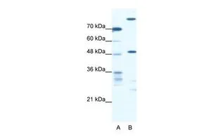
WB analysis of Jurkat cells using GTX47774 NFS1 antibody at 0.3125μg/ml.
Lane A : marker
Lane B : Jurkat cells
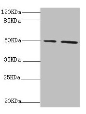
WB analysis of Jurkat cells using GTX47774 NFS1 antibody at 0.3125μg/ml.
Lane A : marker
Lane B : Jurkat cells
NFS1 antibody, Internal
GTX47774
ApplicationsWestern Blot
Product group Antibodies
ReactivityHuman
TargetNFS1
Overview
- SupplierGeneTex
- Product NameNFS1 antibody, Internal
- Delivery Days Customer9
- Application Supplier NoteWB: 0.2-2.5 ug/ml. *Optimal dilutions/concentrations should be determined by the researcher.Not tested in other applications.
- ApplicationsWestern Blot
- CertificationResearch Use Only
- ClonalityPolyclonal
- Concentration0.5-1 mg/ml
- ConjugateUnconjugated
- Gene ID9054
- Target nameNFS1
- Target descriptionNFS1 cysteine desulfurase
- Target synonymsCOXPD52, HUSSY-08, IscS, NIFS, cysteine desulfurase, NFS1 nitrogen fixation 1 homolog, cysteine desulfurase, mitochondrial, nitrogen fixation 1 (S. cerevisiae, homolog), nitrogen-fixing bacteria S-like protein
- HostRabbit
- IsotypeIgG
- Protein IDQ9Y697
- Protein NameCysteine desulfurase
- Scientific DescriptionIron-sulfur clusters are required for the function of many cellular enzymes. The proteins encoded by this gene supply inorganic sulfur to these clusters by removing the sulfur from cysteine, creating alanine in the process. This gene uses alternate in-frame translation initiation sites to generate mitochondrial forms and cytoplasmic/nuclear forms. Selection of the alternative initiation sites is determined by the cytosolic pH. The encoded proteins belong to the class-V family of pyridoxal phosphate-dependent aminotransferases. Alternatively spliced transcript variants have been described. [provided by RefSeq, Nov 2010]
- ReactivityHuman
- Storage Instruction-20°C or -80°C,2°C to 8°C
- UNSPSC41116161
Datasheet
Related products

Product group Antibodies
Anti-NFS1 Antibody Picoband(r)A05061-1-CARRIER-FREE
ApplicationsFlow Cytometry, ImmunoPrecipitation, Western Blot, ELISA
ReactivityHuman, Mouse, Rat
TargetNFS1
- SizePrice

Product group Antibodies
Anti-NFS1 AntibodyA31505
ApplicationsImmunoFluorescence, Western Blot, ImmunoHistoChemistry
ReactivityHuman, Mouse, Rat
- SizePrice
Product group Antibodies

NFS1 AntibodyLS-C830909
ApplicationsELISA, ImmunoHistoChemistry
ReactivityHuman
TargetNFS1
- SizePrice

Product group Antibodies
Anti-NFS1 AntibodyHPA051801
ApplicationsImmunoCytoChemistry, ImmunoHistoChemistry
ReactivityHuman
TargetNFS1
- SizePrice
Product group Antibodies
NFS1 AntibodyCSB-PA896542ESR1HU
ApplicationsImmunoPrecipitation, Western Blot, ELISA, ImmunoHistoChemistry
ReactivityHuman
TargetNFS1
- SizePrice
Product group Antibodies
Nfs1 Polyclonal AntibodyCAC10730
ApplicationsImmunoPrecipitation, Western Blot, ELISA, ImmunoHistoChemistry
TargetNFS1
- SizePrice
Product group Antibodies
References
NFS1 antibodyGTX33356
ApplicationsImmunoFluorescence, Western Blot, ImmunoCytoChemistry, ImmunoHistoChemistry, ImmunoHistoChemistry Paraffin
ReactivityHuman, Mouse, Rat
TargetNFS1
- SizePrice
Product group Antibodies
Anti-NFS1 Antibody144-06668
ApplicationsImmunoFluorescence, Western Blot
ReactivityHuman, Mouse, Rat
TargetNFS1
- SizePrice