

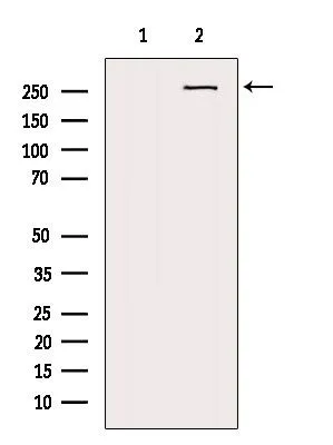
WB analysis of mouse lung tissue lysate using GTX03452 NG2 antibody. The lane on the left was treated with blocking peptide.


WB analysis of mouse lung tissue lysate using GTX03452 NG2 antibody. The lane on the left was treated with blocking peptide.
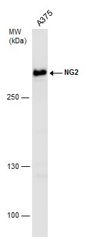
NG2 antibody
GTX03452
ApplicationsImmunoFluorescence, Western Blot, ImmunoCytoChemistry, ImmunoHistoChemistry, ImmunoHistoChemistry Paraffin
Product group Antibodies
ReactivityHuman, Mouse
TargetCSPG4
Overview
- SupplierGeneTex
- Product NameNG2 antibody
- Delivery Days Customer12
- Application Supplier NoteWB: 1:500-1:2000. ICC/IF: 1:100-1:500. IHC-P: 1:50-1:200. *Optimal dilutions/concentrations should be determined by the researcher.Not tested in other applications.
- ApplicationsImmunoFluorescence, Western Blot, ImmunoCytoChemistry, ImmunoHistoChemistry, ImmunoHistoChemistry Paraffin
- CertificationResearch Use Only
- ClonalityPolyclonal
- Concentration1 mg/ml
- ConjugateUnconjugated
- Gene ID1464
- Target nameCSPG4
- Target descriptionchondroitin sulfate proteoglycan 4
- Target synonymsCSPG4A, HMW-MAA, MCSP, MCSPG, MEL-CSPG, MSK16, NG2, chondroitin sulfate proteoglycan 4, chondroitin sulfate proteoglycan 4 (melanoma-associated), chondroitin sulfate proteoglycan NG2, melanoma chondroitin sulfate proteoglycan, melanoma-associated chondroitin sulfate proteoglycan
- HostRabbit
- IsotypeIgG
- Protein IDQ6UVK1
- Protein NameChondroitin sulfate proteoglycan 4
- Scientific DescriptionA human melanoma-associated chondroitin sulfate proteoglycan plays a role in stabilizing cell-substratum interactions during early events of melanoma cell spreading on endothelial basement membranes. CSPG4 represents an integral membrane chondroitin sulfate proteoglycan expressed by human malignant melanoma cells. [provided by RefSeq, Jul 2008]
- ReactivityHuman, Mouse
- Storage Instruction-20°C or -80°C,2°C to 8°C
- UNSPSC41116161
Datasheet
Related products
Product group Antibodies

Anti-MCSP [LC007 (M4-3-ML2)]Ab02805-1.1
ApplicationsFlow Cytometry, ImmunoFluorescence, ELISA, ImmunoHistoChemistry
ReactivityHuman, Monkey
TargetCSPG4
- SizePrice

Product group Antibodies
Anti-NG2/CSPG4 Antibody Picoband(r)A03394-3-CARRIER-FREE
ApplicationsWestern Blot, ELISA
ReactivityHuman
TargetCSPG4
- SizePrice
Product group Antibodies
Anti-CSPG4 Antibody144-03592
ApplicationsWestern Blot
ReactivityHuman
TargetCSPG4
- SizePrice
Product group Antibodies
NG2 Recombinant AntibodyBSM-52891R
ApplicationsWestern Blot
ReactivityHuman
TargetCSPG4
- SizePrice
Product group Antibodies
CSPG4 AntibodyCSB-PA006076ESR1HU
ApplicationsWestern Blot, ELISA, ImmunoHistoChemistry
ReactivityHuman
TargetCSPG4
- SizePrice
Product group Antibodies
ApplicationsImmunoPrecipitation, Western Blot, ImmunoCytoChemistry, ImmunoHistoChemistry
ReactivityRat
TargetCSPG4
- SizePrice

Product group Antibodies
Anti-CSPG4 AntibodyHPA002951
ApplicationsWestern Blot, ImmunoHistoChemistry
ReactivityHuman
TargetCSPG4
- SizePrice
Product group Antibodies
NG2 antibodyGTX130174
ApplicationsWestern Blot
ReactivityHuman
TargetCSPG4
- SizePrice
Product group Antibodies
NG2 antibodyGTX130401
ApplicationsWestern Blot
ReactivityHuman
TargetCSPG4
- SizePrice
![ELISA analysis of antigen using GTX60751 NG2 antibody [7G4E5].
Black : Control antigen 100ng
Purple : Antigen 10ng
Blue : Antigen 50ng
Red : Antigen 100ng](https://www.genetex.com/upload/website/prouct_img/normal/GTX60751/GTX60751_20170912_ELISA_w_23061123_764.webp)
Product group Antibodies
NG2 antibody [7G4E5]GTX60751
ApplicationsFlow Cytometry, Western Blot, ELISA
ReactivityHuman
TargetCSPG4
- SizePrice