

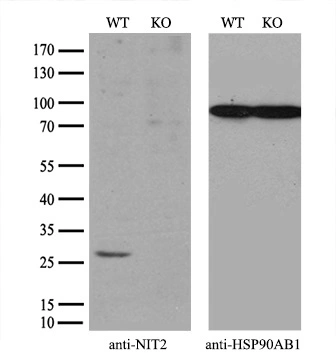
WB analysis of wild type and NIT2 knock out 293T cell lysate(10μg per lane) using GTX84040 NIT2 antibody [6H7]. Dilution : 1:100
![IHC-P analysis of kidney tissue using GTX84040 NIT2 antibody [6H7]. Antigen retrieval : Heat-induced epitope retrieval by 10mM citrate buffer, pH6.0, 100oC for 10min. Dilution : 1:50](https://www.genetex.com/upload/website/prouct_img/normal/GTX84040/GTX84040_2226_IHC-P_w_23061420_448.webp "IHC-P analysis of kidney tissue using GTX84040 NIT2 antibody [6H7]. Antigen retrieval : Heat-induced epitope retrieval by 10mM citrate buffer, pH6.0, 100oC for 10min. Dilution : 1:50")
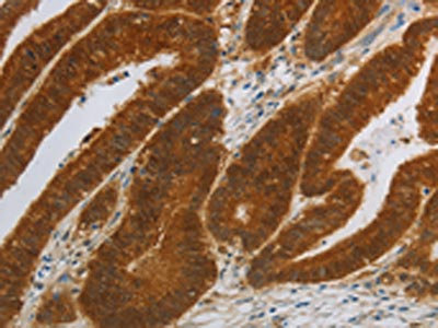
![IHC-P analysis of colon tissue using GTX84040 NIT2 antibody [6H7]. Antigen retrieval : Heat-induced epitope retrieval by 10mM citrate buffer, pH6.0, 100oC for 10min. Dilution : 1:50](https://www.genetex.com/upload/website/prouct_img/normal/GTX84040/GTX84040_2225_IHC-P_w_23061420_808.webp "IHC-P analysis of colon tissue using GTX84040 NIT2 antibody [6H7]. Antigen retrieval : Heat-induced epitope retrieval by 10mM citrate buffer, pH6.0, 100oC for 10min. Dilution : 1:50")
![IHC-P analysis of colon adenocarcinoma tissue using GTX84040 NIT2 antibody [6H7]. Antigen retrieval : Heat-induced epitope retrieval by 10mM citrate buffer, pH6.0, 100oC for 10min. Dilution : 1:50](https://www.genetex.com/upload/website/prouct_img/normal/GTX84040/GTX84040_2223_IHC-P_w_23061420_820.webp "IHC-P analysis of colon adenocarcinoma tissue using GTX84040 NIT2 antibody [6H7]. Antigen retrieval : Heat-induced epitope retrieval by 10mM citrate buffer, pH6.0, 100oC for 10min. Dilution : 1:50")
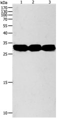
![WB analysis of HEK293T cells transfected with NIT2 plasmid (Right) or empty vector (Left) for 48 hrs using GTX84040 NIT2 antibody [6H7]. Loading : 5 ug per lane](https://www.genetex.com/upload/website/prouct_img/normal/GTX84040/GTX84040_4086_WB_w_23061420_719.webp "WB analysis of HEK293T cells transfected with NIT2 plasmid (Right) or empty vector (Left) for 48 hrs using GTX84040 NIT2 antibody [6H7]. Loading : 5 ug per lane")
![IHC-P analysis of prostate carcinoma tissue using GTX84040 NIT2 antibody [6H7]. Antigen retrieval : Heat-induced epitope retrieval by 10mM citrate buffer, pH6.0, 100oC for 10min. Dilution : 1:50](https://www.genetex.com/upload/website/prouct_img/normal/GTX84040/GTX84040_2224_IHC-P_w_23061420_722.webp "IHC-P analysis of prostate carcinoma tissue using GTX84040 NIT2 antibody [6H7]. Antigen retrieval : Heat-induced epitope retrieval by 10mM citrate buffer, pH6.0, 100oC for 10min. Dilution : 1:50")
![ICC/IF analysis of COS7 cells transiently transfected with NIT2 plasmid using GTX84040 NIT2 antibody [6H7].](https://www.genetex.com/upload/website/prouct_img/normal/GTX84040/GTX84040_922_ICCIF_w_23061420_114.webp "ICC/IF analysis of COS7 cells transiently transfected with NIT2 plasmid using GTX84040 NIT2 antibody [6H7].")
WB analysis of wild type and NIT2 knock out 293T cell lysate(10μg per lane) using GTX84040 NIT2 antibody [6H7]. Dilution : 1:100
NIT2 antibody [6H7]
GTX84040
ApplicationsImmunoFluorescence, Western Blot, ImmunoCytoChemistry, ImmunoHistoChemistry, ImmunoHistoChemistry Paraffin
Product group Antibodies
ReactivityHuman
TargetNIT2
Overview
- SupplierGeneTex
- Product NameNIT2 antibody [6H7]
- Delivery Days Customer9
- Application Supplier NoteWB: 1:1000. ICC/IF: 1:100. IHC-P: 1:50. *Optimal dilutions/concentrations should be determined by the researcher.Not tested in other applications.
- ApplicationsImmunoFluorescence, Western Blot, ImmunoCytoChemistry, ImmunoHistoChemistry, ImmunoHistoChemistry Paraffin
- CertificationResearch Use Only
- ClonalityMonoclonal
- Clone ID6H7
- Concentration0.71 mg/ml
- ConjugateUnconjugated
- Gene ID56954
- Target nameNIT2
- Target descriptionnitrilase family member 2
- Target synonymsHEL-S-8a, omega-amidase NIT2, Nit protein 2, epididymis secretory sperm binding protein Li 8a, nitrilase homolog 2
- HostMouse
- IsotypeIgG2b
- Protein IDQ9NQR4
- Protein NameOmega-amidase NIT2
- Scientific DescriptionHas a omega-amidase activity. The role of omega-amidase is to remove potentially toxic intermediates by converting alpha-ketoglutaramate and alpha-ketosuccinamate to biologically useful alpha-ketoglutarate and oxaloacetate, respectively. Overexpression decreases the colony-forming capacity of cultured cells by arresting cells in the G2 phase of the cell cycle.
- ReactivityHuman
- Storage Instruction-20°C or -80°C,2°C to 8°C
- UNSPSC41116161
Datasheet
Related products
Product group Antibodies
NIT2 AntibodyCSB-PA187627
ApplicationsWestern Blot, ELISA, ImmunoHistoChemistry
ReactivityHuman, Mouse
TargetNIT2
- SizePrice

Product group Antibodies
Anti-NIT2 Antibody Picoband(r)A05891-3-CARRIER-FREE
ApplicationsFlow Cytometry, Western Blot, ELISA, ImmunoHistoChemistry
ReactivityHuman, Mouse, Rat
TargetNIT2
- SizePrice
Product group Antibodies
Anti-NIT2 AntibodyA48501
ApplicationsWestern Blot, ELISA, ImmunoHistoChemistry
ReactivityHuman, Mouse
- SizePrice

Product group Antibodies
Anti-NIT2 AntibodyHPA036999
ApplicationsWestern Blot, ImmunoCytoChemistry, ImmunoHistoChemistry
ReactivityHuman, Mouse, Rat
TargetNIT2
- SizePrice
Product group Antibodies

NIT2 AntibodyLS-C405959
ApplicationsWestern Blot, ELISA, ImmunoHistoChemistry
ReactivityHuman, Mouse
TargetNIT2
- SizePrice
![FACS analysis of HEK293T cells transfected with either NIT2 plasmid(Red) or empty vector control plasmid(Blue) using GTX84036 NIT2 antibody [2B9].](https://www.genetex.com/upload/website/prouct_img/normal/GTX84036/GTX84036_281_FACS_w_23061420_986.webp)
Product group Antibodies
NIT2 antibody [2B9]GTX84036
ApplicationsFlow Cytometry, ImmunoFluorescence, Western Blot, ImmunoCytoChemistry, ImmunoHistoChemistry, ImmunoHistoChemistry Paraffin
ReactivityHuman
TargetNIT2
- SizePrice
![IHC-P analysis of colon adenocarcinoma tissue using GTX84039 NIT2 antibody [3D5]. Antigen retrieval : Heat-induced epitope retrieval by 10mM citrate buffer, pH6.0, 100oC for 10min. Dilution : 1:50](https://www.genetex.com/upload/website/prouct_img/normal/GTX84039/GTX84039_2220_IHC-P_w_23061420_446.webp)
Product group Antibodies
NIT2 antibody [3D5]GTX84039
ApplicationsFlow Cytometry, ImmunoFluorescence, Western Blot, ImmunoCytoChemistry, ImmunoHistoChemistry, ImmunoHistoChemistry Paraffin
ReactivityHuman, Monkey, Mouse, Rat
TargetNIT2
- SizePrice
Product group Antibodies
NIT2 Polyclonal AntibodyCAC14456
ApplicationsWestern Blot, ELISA
ReactivityMouse
TargetNIT2
- SizePrice