

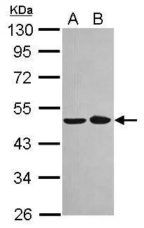
Sample (30 ug of whole cell lysate) A: A431 B: Jurkat 10% SDS PAGE GTX120935 diluted at 1:1000
Sample (30 ug of whole cell lysate) A: A431 B: Jurkat 10% SDS PAGE GTX120935 diluted at 1:1000
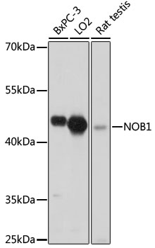
NOB1 antibody [N1N3]
GTX120935
ApplicationsWestern Blot
Product group Antibodies
ReactivityHuman
TargetNOB1
Overview
- SupplierGeneTex
- Product NameNOB1 antibody [N1N3]
- Delivery Days Customer9
- Application Supplier NoteWB: 1:500-1:3000. *Optimal dilutions/concentrations should be determined by the researcher.Not tested in other applications.
- ApplicationsWestern Blot
- CertificationResearch Use Only
- ClonalityPolyclonal
- Concentration1.01 mg/ml
- ConjugateUnconjugated
- Gene ID28987
- Target nameNOB1
- Target descriptionNIN1 (RPN12) binding protein 1 homolog
- Target synonymsART-4, MST158, MSTP158, NOB1P, PSMD8BP1, RNA-binding protein NOB1, NIN1/PSMD8 binding protein 1 homolog, PSMD8 binding protein 1, adenocarcinoma antigen recognized by T lymphocytes 4, nin one binding protein, phosphorylation regulatory protein HP-10, protein ART-4
- HostRabbit
- IsotypeIgG
- Protein IDQ9ULX3
- Protein NameRNA-binding protein NOB1
- Scientific DescriptionIn yeast, over 200 protein and RNA cofactors are required for ribosome assembly, and these are generally conserved in eukaryotes. These factors orchestrate modification and cleavage of the initial 35S precursor rRNA transcript into the mature 18S, 5.8S, and 25S rRNAs, folding of the rRNA, and binding of ribosomal proteins and 5S RNA. Nob1 is involved in pre-rRNA processing. In a late cytoplasmic processing step, Nob1 cleaves a 20S rRNA intermediate at cleavage site D to produce the mature 18S rRNA (Lamanna and Karbstein, 2009 [PubMed 19706509]).[supplied by OMIM]
- ReactivityHuman
- Storage Instruction-20°C or -80°C,2°C to 8°C
- UNSPSC41116161
Datasheet
Related products

Product group Antibodies
Anti-NOB1 Antibody Picoband(r)A03504-1-CARRIER-FREE
ApplicationsWestern Blot, ELISA, ImmunoHistoChemistry
ReactivityHuman, Mouse, Rat
TargetNOB1
- SizePrice
Product group Antibodies
Anti-NOB1 Antibody144-65083
ApplicationsImmunoFluorescence, Western Blot
ReactivityHuman, Mouse, Rat
TargetNOB1
- SizePrice
Product group Antibodies
Anti-NOB1 AntibodyA90093
ApplicationsImmunoFluorescence, Western Blot, ImmunoCytoChemistry
ReactivityHuman, Mouse, Rat
- SizePrice
Product group Antibodies
NOB1 Polyclonal AntibodyBS-8857R
ApplicationsImmunoFluorescence, Western Blot, ImmunoHistoChemistry, ImmunoHistoChemistry Paraffin
ReactivityHuman, Mouse, Rat
TargetNOB1
- SizePrice
Product group Antibodies
NOB1 AntibodyCSB-PA892472LA01HU
ApplicationsImmunoFluorescence, Western Blot, ELISA, ImmunoHistoChemistry
ReactivityHuman, Mouse
TargetNOB1
- SizePrice
Product group Antibodies
Nob1 Polyclonal AntibodyCAC07953
ApplicationsImmunoFluorescence, Western Blot, ELISA, ImmunoHistoChemistry
ReactivityMouse
TargetNOB1
- SizePrice
Product group Antibodies

NOB1 / NOB1P AntibodyLS-C502160
ApplicationsImmunoFluorescence, Western Blot, ELISA, ImmunoHistoChemistry
ReactivityHuman, Mouse
TargetNOB1
- SizePrice
Product group Antibodies
Anti-NOB1 AntibodyHPA049430
ApplicationsImmunoHistoChemistry
ReactivityHuman
TargetNOB1
- SizePrice