

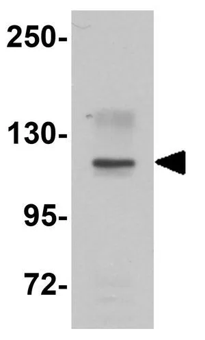
WB analysis of EL4 cell lysate using GTX31810 NOD1 antibody. Working concentration : 1 μg/ml
WB analysis of EL4 cell lysate using GTX31810 NOD1 antibody. Working concentration : 1 μg/ml
NOD1 antibody
GTX31810
ApplicationsWestern Blot, ELISA
Product group Antibodies
ReactivityHuman, Mouse, Rat
TargetNOD1
Overview
- SupplierGeneTex
- Product NameNOD1 antibody
- Delivery Days Customer9
- Application Supplier NoteWB: 1 microg/mL. *Optimal dilutions/concentrations should be determined by the researcher.Not tested in other applications.
- ApplicationsWestern Blot, ELISA
- CertificationResearch Use Only
- ClonalityPolyclonal
- Concentration1 mg/ml
- ConjugateUnconjugated
- Gene ID10392
- Target nameNOD1
- Target descriptionnucleotide binding oligomerization domain containing 1
- Target synonymsCARD4, CLR7.1, NLRC1, hNod1, nucleotide-binding oligomerization domain-containing protein 1, NLR family, CARD domain containing 1, caspase recruitment domain family, member 4, caspase recruitment domain-containing protein 4, nucleotide-binding oligomerization domain, leucine rich repeat and CARD domain containing 1
- HostRabbit
- IsotypeIgG
- Protein IDQ9Y239
- Protein NameNucleotide-binding oligomerization domain-containing protein 1
- Scientific DescriptionThis gene encodes a member of the NOD (nucleotide-binding oligomerization domain) family. This member is a cytosolic protein. It contains an N-terminal caspase recruitment domain (CARD), a centrally located nucleotide-binding domain (NBD), and 10 tandem leucine-rich repeats (LRRs) in its C terminus. The CARD is involved in apoptotic signaling, LRRs participate in protein-protein interactions, and mutations in the NBD may affect the process of oligomerization and subsequent function of the LRR domain. This protein is an intracellular pattern-recognition receptor (PRR) that initiates inflammation in response to a subset of bacteria through the detection of bacterial diaminopimelic acid. Multiple alternatively spliced transcript variants differring in the 5 UTR have been described, but the full-length nature of these variants has not been determined. [provided by RefSeq, Oct 2009]
- ReactivityHuman, Mouse, Rat
- Storage Instruction-20°C or -80°C,2°C to 8°C
- UNSPSC41116161
Datasheet
Related products
Product group Antibodies
Anti-NOD1 Antibody144-62342
ApplicationsWestern Blot, ImmunoHistoChemistry
ReactivityHuman, Mouse, Rat
TargetNOD1
- SizePrice
Product group Antibodies
Anti-NOD1 AntibodyA13404
ApplicationsWestern Blot, ImmunoHistoChemistry
ReactivityHuman, Mouse, Rat
- SizePrice
Product group Antibodies
References
CARD4 Polyclonal AntibodyBS-7085R
ApplicationsImmunoFluorescence, Western Blot, ELISA, ImmunoCytoChemistry, ImmunoHistoChemistry, ImmunoHistoChemistry Frozen, ImmunoHistoChemistry Paraffin
ReactivityHuman, Mouse, Rat
TargetNOD1
- SizePrice

Product group Antibodies
Anti-CARD4/NOD1 Antibody Picoband(r)A00495-1-CARRIER-FREE
ApplicationsWestern Blot
ReactivityMonkey
TargetNOD1
- SizePrice
Product group Antibodies

Goat anti-NOD1 / CARD4EB05389
ApplicationsWestern Blot, ELISA
ReactivityBovine, Canine, Human, Mouse, Porcine, Rat
TargetNOD1
- SizePrice
Product group Antibodies
NOD1 AntibodyCSB-PA047815
ApplicationsELISA, ImmunoHistoChemistry
ReactivityHuman, Mouse
TargetNOD1
- SizePrice
Product group Antibodies
ApplicationsImmunoPrecipitation, Western Blot, ImmunoCytoChemistry, ImmunoHistoChemistry
TargetNOD1
- SizePrice
Product group Antibodies

NOD1 AntibodyLS-C403024
ApplicationsELISA, ImmunoHistoChemistry
ReactivityHuman, Mouse
TargetNOD1
- SizePrice

Product group Antibodies
Anti-NOD1 AntibodyHPA074367
ApplicationsImmunoCytoChemistry
ReactivityHuman
TargetNOD1
- SizePrice
![NOD1 antibody [C2C3], C-term detects NOD1 protein by Western blot analysis. Various whole cell extracts (30 μg) were separated by 7.5% SDS-PAGE, and the membrane was blotted with NOD1 antibody [C2C3], C-term (GTX108811) diluted at a dilution of 1:500.](https://www.genetex.com/upload/website/prouct_img/normal/GTX108811/GTX108811_41766_20150605_WB_22090701_155.webp)
Product group Antibodies
NOD1 antibody [C2C3], C-termGTX108811
ApplicationsImmunoFluorescence, Western Blot, ImmunoCytoChemistry
ReactivityHuman
TargetNOD1
- SizePrice