

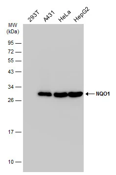
Various whole cell extracts (30 μg) were separated by 12% SDS-PAGE, and the membrane was blotted with NQO1 antibody [C2C3], C-term (GTX100235) diluted at 1:1000. The HRP-conjugated anti-rabbit IgG antibody (GTX213110-01) was used to detect the primary antibody.
 antibody at 1:200 dilution.")
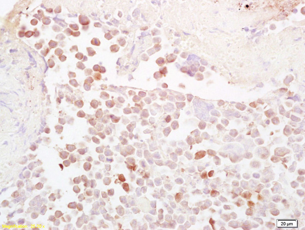
![NQO1 antibody [C2C3], C-term detects NQO1 protein at cytoplasm in human rectal carcinoma by immunohistochemical analysis. Sample: Paraffin-embedded human rectal carcinoma. NQO1 antibody [C2C3], C-term (GTX100235) diluted at 1:500.
Antigen Retrieval: Trilogy? (EDTA based, pH 8.0) buffer, 15min](https://www.genetex.com/upload/website/prouct_img/normal/GTX100235/GTX100235_40073_20160125_IHC-P_w_23053123_934.webp "NQO1 antibody [C2C3], C-term detects NQO1 protein at cytoplasm in human rectal carcinoma by immunohistochemical analysis. Sample: Paraffin-embedded human rectal carcinoma. NQO1 antibody [C2C3], C-term (GTX100235) diluted at 1:500.
Antigen Retrieval: Trilogy? (EDTA based, pH 8.0) buffer, 15min")
![NQO1 antibody [C2C3], C-term detects NQO1 protein at cytoplasm by immunohistochemical analysis. Sample: Paraffin-embedded mouse kidney. NQO1 stained by NQO1 antibody [C2C3], C-term (GTX100235) diluted at 1:500. Antigen Retrieval: Citrate buffer, pH 6.0, 15 min](https://www.genetex.com/upload/website/prouct_img/normal/GTX100235/GTX100235_43146_20200122_IHC-P_M_w_23053123_298.webp "NQO1 antibody [C2C3], C-term detects NQO1 protein at cytoplasm by immunohistochemical analysis. Sample: Paraffin-embedded mouse kidney. NQO1 stained by NQO1 antibody [C2C3], C-term (GTX100235) diluted at 1:500. Antigen Retrieval: Citrate buffer, pH 6.0, 15 min")
![NQO1 antibody [C2C3], C-term detects NQO1 protein by western blot analysis. A. 50 μg mouse heart lysate/extract 12% SDS-PAGE NQO1 antibody [C2C3], C-term (GTX100235) dilution: 1:500 The HRP-conjugated anti-rabbit IgG antibody (GTX213110-01) was used to detect the primary antibody.](https://www.genetex.com/upload/website/prouct_img/normal/GTX100235/GTX100235_40073_WB_M_heart_w_23053123_325.webp "NQO1 antibody [C2C3], C-term detects NQO1 protein by western blot analysis. A. 50 μg mouse heart lysate/extract 12% SDS-PAGE NQO1 antibody [C2C3], C-term (GTX100235) dilution: 1:500 The HRP-conjugated anti-rabbit IgG antibody (GTX213110-01) was used to detect the primary antibody.")
![NQO1 antibody [C2C3], C-term detects NQO1 protein by western blot analysis. A. 50 μg rat heart lysate/extract 12% SDS-PAGE NQO1 antibody [C2C3], C-term (GTX100235) dilution: 1:500 The HRP-conjugated anti-rabbit IgG antibody (GTX213110-01) was used to detect the primary antibody.](https://www.genetex.com/upload/website/prouct_img/normal/GTX100235/GTX100235_40073_WB_R_heart_w_23053123_400.webp "NQO1 antibody [C2C3], C-term detects NQO1 protein by western blot analysis. A. 50 μg rat heart lysate/extract 12% SDS-PAGE NQO1 antibody [C2C3], C-term (GTX100235) dilution: 1:500 The HRP-conjugated anti-rabbit IgG antibody (GTX213110-01) was used to detect the primary antibody.")
Various whole cell extracts (30 μg) were separated by 12% SDS-PAGE, and the membrane was blotted with NQO1 antibody [C2C3], C-term (GTX100235) diluted at 1:1000. The HRP-conjugated anti-rabbit IgG antibody (GTX213110-01) was used to detect the primary antibody.
NQO1 antibody [C2C3], C-term
GTX100235
ApplicationsImmunoFluorescence, Western Blot, ImmunoCytoChemistry, ImmunoHistoChemistry, ImmunoHistoChemistry Paraffin
Product group Antibodies
ReactivityHuman, Monkey, Mouse, Rat
TargetNQO1
Overview
- SupplierGeneTex
- Product NameNQO1 antibody [C2C3], C-term
- Delivery Days Customer9
- Application Supplier NoteWB: 1:500-1:3000. ICC/IF: 1:100-1:1000. IHC-P: 1:100-1:1000. *Optimal dilutions/concentrations should be determined by the researcher.Not tested in other applications.
- ApplicationsImmunoFluorescence, Western Blot, ImmunoCytoChemistry, ImmunoHistoChemistry, ImmunoHistoChemistry Paraffin
- CertificationResearch Use Only
- ClonalityPolyclonal
- Concentration0.61 mg/ml
- ConjugateUnconjugated
- Gene ID1728
- Target nameNQO1
- Target descriptionNAD(P)H quinone dehydrogenase 1
- Target synonymsDHQU, DIA4, DTD, NMOR1, NMORI, QR1, NAD(P)H dehydrogenase [quinone] 1, DT-diaphorase, NAD(P)H dehydrogenase, quinone 1, NAD(P)H-quinone oxidoreductase, NAD(P)H:Quinone acceptor oxidoreductase type 1, NAD(P)H:menadione oxidoreductase 1, NAD(P)H:quinone oxidoreductase 1, NAD(P)H:quinone oxireductase, azoreductase, diaphorase (NADH/NADPH) (cytochrome b-5 reductase), diaphorase-4, dioxin-inducible 1, menadione reductase, phylloquinone reductase, quinone reductase 1
- HostRabbit
- IsotypeIgG
- Protein IDP15559
- Protein NameNAD(P)H dehydrogenase [quinone] 1
- Scientific DescriptionThis gene is a member of the NAD(P)H dehydrogenase (quinone) family and encodes a cytoplasmic 2-electron reductase. This FAD-binding protein forms homodimers and reduces quinones to hydroquinones. This proteins enzymatic activity prevents the one electron reduction of quinones that results in the production of radical species. Mutations in this gene have been associated with tardive dyskinesia (TD), an increased risk of hematotoxicity after exposure to benzene, and susceptibility to various forms of cancer. Altered expression of this protein has been seen in many tumors and is also associated with Alzheimers disease (AD). Alternate transcriptional splice variants, encoding different isoforms, have been characterized. [provided by RefSeq]
- ReactivityHuman, Monkey, Mouse, Rat
- Storage Instruction-20°C or -80°C,2°C to 8°C
- UNSPSC41116161
Datasheet
Related products
Product group Antibodies
NQO1 AntibodyCSB-PA003475
ApplicationsWestern Blot, ELISA
ReactivityHuman
TargetNQO1
- SizePrice

Product group Antibodies
Anti-NQO1 Antibody Picoband(r)A00494-2-CARRIER-FREE
ApplicationsFlow Cytometry, ImmunoFluorescence, Western Blot, ELISA, ImmunoCytoChemistry
ReactivityHuman
TargetNQO1
- SizePrice
Product group Antibodies
Anti-NQO1 AntibodyA101354
ApplicationsWestern Blot, ELISA
ReactivityHuman
- SizePrice
Product group Antibodies

Goat anti-NQO1EB05370
ApplicationsImmunoFluorescence, Western Blot, ELISA, ImmunoHistoChemistry
ReactivityCanine, Human, Porcine, Rat
TargetNQO1
- SizePrice

Product group Antibodies
Anti-NQO1 AntibodyHPA007308
ApplicationsWestern Blot, ImmunoCytoChemistry, ImmunoHistoChemistry
ReactivityHuman
TargetNQO1
- SizePrice
Product group Antibodies

NQO1 AntibodyLS-C405968
ApplicationsWestern Blot, ELISA, ImmunoHistoChemistry
ReactivityHuman, Mouse, Rat
TargetNQO1
- SizePrice
![IHC-P analysis of human breast carcinoma tissue using GTX30626 NQO1 antibody [A180].](https://www.genetex.com/upload/website/prouct_img/normal/GTX30626/GTX30626_562_IHC-P_w_23060722_691.webp)
Product group Antibodies
References
NQO1 antibody [A180]GTX30626
ApplicationsFlow Cytometry, ImmunoFluorescence, ImmunoPrecipitation, Western Blot, ImmunoCytoChemistry, ImmunoHistoChemistry, ImmunoHistoChemistry Frozen, ImmunoHistoChemistry Paraffin
ReactivityCanine, Human, Mouse, Primate, Rat
TargetNQO1
- SizePrice
Product group Antibodies
ApplicationsImmunoPrecipitation, Western Blot, ImmunoCytoChemistry, ImmunoHistoChemistry
ReactivityMouse, Rat
TargetNQO1
- SizePrice
Product group Antibodies
References
NQO1 Polyclonal AntibodyBS-2184R
ApplicationsImmunoFluorescence, Western Blot, ELISA, ImmunoCytoChemistry, ImmunoHistoChemistry, ImmunoHistoChemistry Frozen, ImmunoHistoChemistry Paraffin
ReactivityEquine, Guinea Pig, Human, Mouse, Porcine, Rabbit, Rat
TargetNQO1
- SizePrice