

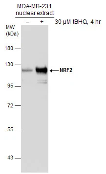
Untreated (–) and treated (+) MDA-MB-231 nuclear extracts (30 μg) were separated by 7.5% SDS-PAGE, and the membrane was blotted with NRF2 antibody [N2C2], Internal (GTX103322) diluted at 1:1000.
![NRF2 antibody [N2C2], Internal detects NRF2 protein at cytoplasm and nucleus by immunofluorescent analysis. Sample: NIH/3T3 cells were fixed in 4% paraformaldehyde at RT for 15 min. Green: NRF2 protein stained by NRF2 antibody [N2C2], Internal (GTX103322) diluted at 1:500. Blue: Hoechst 33342 staining. Scale bar = 10 μm.](https://www.genetex.com/upload/website/prouct_img/normal/GTX103322/GTX103322_42004_20160720_IFA_w_23060119_806.webp "NRF2 antibody [N2C2], Internal detects NRF2 protein at cytoplasm and nucleus by immunofluorescent analysis. Sample: NIH/3T3 cells were fixed in 4% paraformaldehyde at RT for 15 min. Green: NRF2 protein stained by NRF2 antibody [N2C2], Internal (GTX103322) diluted at 1:500. Blue: Hoechst 33342 staining. Scale bar = 10 μm.")
![Untreated (–) and treated (+) HepG2 whole cell extracts (30 μg) were separated by 5% SDS-PAGE, and the membranes were blotted with NRF2 antibody [N2C2], Internal (GTX103322) diluted at 1:500 and competitor's antibody diluted at 1:500. The HRP-conjugated anti-rabbit IgG antibody (GTX213110-01) was used to detect the primary antibody. *The competitor is not affiliated with GeneTex and does not endorse this product.](https://www.genetex.com/upload/website/prouct_img/normal/GTX103322/GTX103322_43552_20200117_WB_treatment_MG132_competitor_watermark_w_23060119_423.webp "Untreated (–) and treated (+) HepG2 whole cell extracts (30 μg) were separated by 5% SDS-PAGE, and the membranes were blotted with NRF2 antibody [N2C2], Internal (GTX103322) diluted at 1:500 and competitor's antibody diluted at 1:500. The HRP-conjugated anti-rabbit IgG antibody (GTX213110-01) was used to detect the primary antibody. *The competitor is not affiliated with GeneTex and does not endorse this product.")
 and NRF2-transfected (+, including 3xFlag-tag) 293T whole cell extracts (30 μg) were separated by 5% SDS-PAGE, and the membrane was blotted with NRF2 antibody (GTX103322) diluted by 1:1000. The HRP-conjugated anti-rabbit IgG antibody (GTX213110-01) was used to detect the primary antibody.")
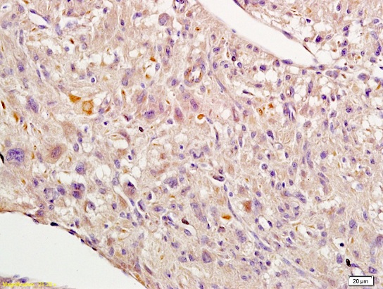
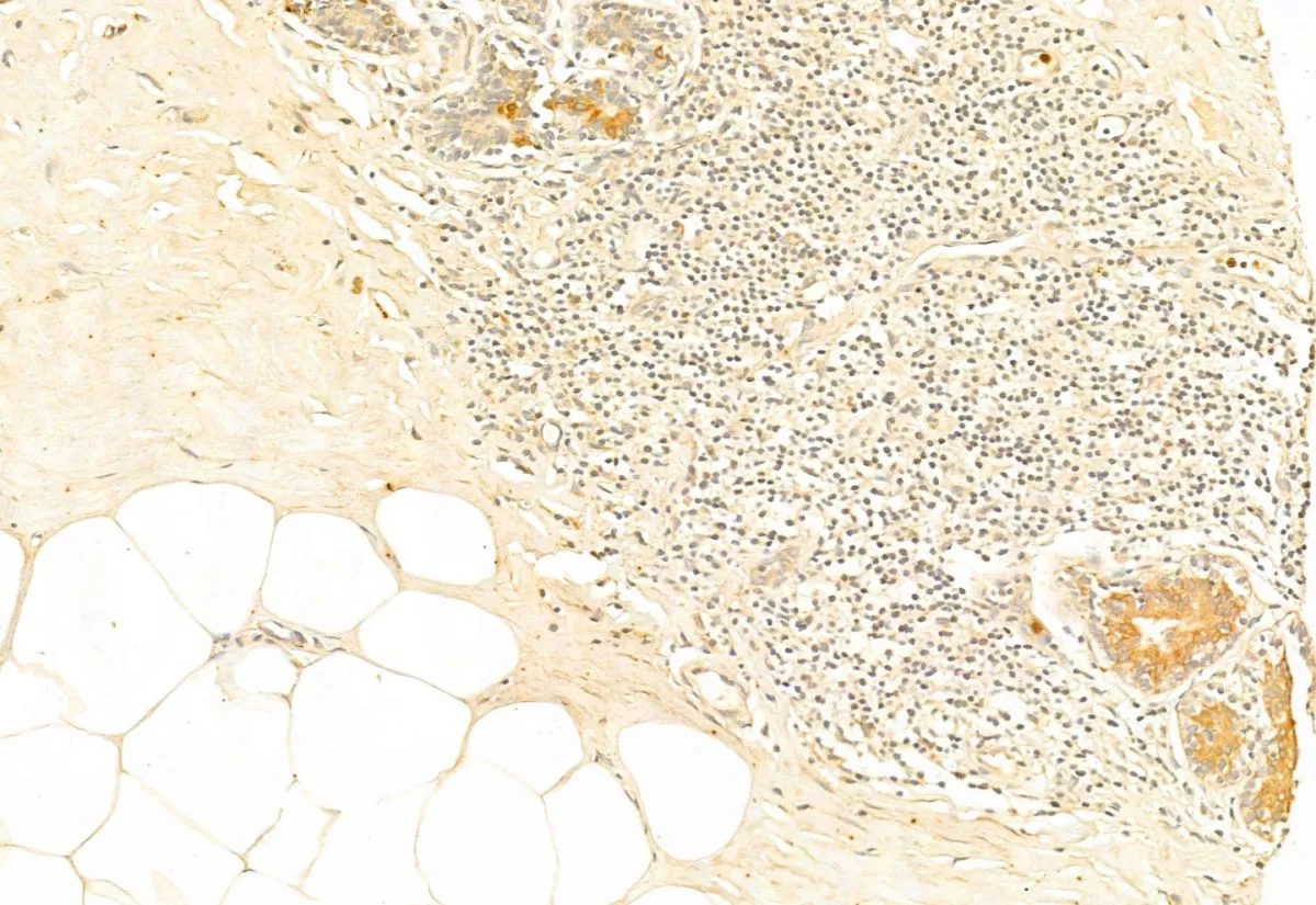
![NRF2 antibody [N2C2], Internal detects NRF2 protein at cytoplasm and nucleus by immunohistochemical analysis. Sample: Paraffin-embedded human breast carcinoma. NRF2 stained by NRF2 antibody [N2C2], Internal (GTX103322) diluted at 1:500. Antigen Retrieval: Citrate buffer, pH 6.0, 15 min](https://www.genetex.com/upload/website/prouct_img/normal/GTX103322/GTX103322_44111_20201127_IHC-P_w_23060119_749.webp "NRF2 antibody [N2C2], Internal detects NRF2 protein at cytoplasm and nucleus by immunohistochemical analysis. Sample: Paraffin-embedded human breast carcinoma. NRF2 stained by NRF2 antibody [N2C2], Internal (GTX103322) diluted at 1:500. Antigen Retrieval: Citrate buffer, pH 6.0, 15 min")

![NRF2 antibody [N2C2], Internal detects NRF2 protein at nucleus by immunofluorescent analysis. Sample: Neuro2A cells were fixed in 4% paraformaldehyde at RT for 15 min. Green: NRF2 stained by NRF2 antibody [N2C2], Internal (GTX103322) diluted at 1:1000.](https://www.genetex.com/upload/website/prouct_img/normal/GTX103322/GTX103322_43677_20210122_ICC_IF_w_23060119_402.webp "NRF2 antibody [N2C2], Internal detects NRF2 protein at nucleus by immunofluorescent analysis. Sample: Neuro2A cells were fixed in 4% paraformaldehyde at RT for 15 min. Green: NRF2 stained by NRF2 antibody [N2C2], Internal (GTX103322) diluted at 1:1000.")
![NRF2 antibody [N2C2], Internal detects NRF2 protein at nucleus by immunofluorescent analysis. Sample: Mock and treated HeLa cells were fixed in 4% paraformaldehyde at RT for 15 min. Green: NRF2 stained by NRF2 antibody [N2C2], Internal (GTX103322) diluted at 1:500. Blue: Fluoroshield with DAPI (GTX30920).](https://www.genetex.com/upload/website/prouct_img/normal/GTX103322/GTX103322_44020_20210924_ICC_IF_treatment_MG132_w_23060119_837.webp "NRF2 antibody [N2C2], Internal detects NRF2 protein at nucleus by immunofluorescent analysis. Sample: Mock and treated HeLa cells were fixed in 4% paraformaldehyde at RT for 15 min. Green: NRF2 stained by NRF2 antibody [N2C2], Internal (GTX103322) diluted at 1:500. Blue: Fluoroshield with DAPI (GTX30920).")
![NRF2 antibody [N2C2], Internal detects NRF2 protein at nucleus by immunofluorescent analysis. Sample: HeLa cells were fixed in 4% paraformaldehyde at RT for 15 min. Green: NRF2 stained by NRF2 antibody [N2C2], Internal (GTX103322) diluted at 1:1000. Red: phalloidin, a cytoskeleton marker, diluted at 1:200. Scale bar= 10 μm.](https://www.genetex.com/upload/website/prouct_img/normal/GTX103322/GTX103322_43636_20200311_ICC_IF_w_23060119_425.webp "NRF2 antibody [N2C2], Internal detects NRF2 protein at nucleus by immunofluorescent analysis. Sample: HeLa cells were fixed in 4% paraformaldehyde at RT for 15 min. Green: NRF2 stained by NRF2 antibody [N2C2], Internal (GTX103322) diluted at 1:1000. Red: phalloidin, a cytoskeleton marker, diluted at 1:200. Scale bar= 10 μm.")
![Untreated (–) and treated (+) RAW264.7 whole cell extracts (30 μg) were separated by 5% SDS-PAGE, and the membrane was blotted with NRF2 antibody [N2C2], Internal (GTX103322) diluted at 1:500. The HRP-conjugated anti-rabbit IgG antibody (GTX213110-01) was used to detect the primary antibody.](https://www.genetex.com/upload/website/prouct_img/normal/GTX103322/GTX103322_43054_20190118_WB_M_treatment_LPS_w_23060119_100.webp "Untreated (–) and treated (+) RAW264.7 whole cell extracts (30 μg) were separated by 5% SDS-PAGE, and the membrane was blotted with NRF2 antibody [N2C2], Internal (GTX103322) diluted at 1:500. The HRP-conjugated anti-rabbit IgG antibody (GTX213110-01) was used to detect the primary antibody.")
Untreated (–) and treated (+) MDA-MB-231 nuclear extracts (30 μg) were separated by 7.5% SDS-PAGE, and the membrane was blotted with NRF2 antibody [N2C2], Internal (GTX103322) diluted at 1:1000.
NRF2 antibody [N2C2], Internal
GTX103322
ApplicationsFlow Cytometry, ImmunoFluorescence, ImmunoPrecipitation, Western Blot, ChIP Chromatin ImmunoPrecipitation, ImmunoCytoChemistry, ImmunoHistoChemistry, ImmunoHistoChemistry Paraffin
Product group Antibodies
ReactivityAvian, Human, Mouse, Plant, Reptile, Rat, Zebra Fish
TargetNFE2L2
Overview
- SupplierGeneTex
- Product NameNRF2 antibody [N2C2], Internal
- Delivery Days Customer9
- Application Supplier NoteWB: 1:500-1:3000. ICC/IF: 1:100-1:1000. IHC-P: 1:100-1:1000. IP: 1:100-1:500. *Optimal dilutions/concentrations should be determined by the researcher.Not tested in other applications.
- ApplicationsFlow Cytometry, ImmunoFluorescence, ImmunoPrecipitation, Western Blot, ChIP Chromatin ImmunoPrecipitation, ImmunoCytoChemistry, ImmunoHistoChemistry, ImmunoHistoChemistry Paraffin
- CertificationResearch Use Only
- ClonalityPolyclonal
- Concentration0.26 mg/ml
- ConjugateUnconjugated
- Gene ID4780
- Target nameNFE2L2
- Target descriptionNFE2 like bZIP transcription factor 2
- Target synonymsHEBP1, IMDDHH, NRF2, Nrf-2, nuclear factor erythroid 2-related factor 2, nuclear factor erythroid-derived 2-like 2, nuclear factor, erythroid 2 like 2
- HostRabbit
- IsotypeIgG
- Protein IDQ16236
- Protein NameNuclear factor erythroid 2-related factor 2
- Scientific DescriptionNFE2 (MIM 601490), NFE2L1 (MIM 163260), and NFE2L2 comprise a family of human genes encoding basic leucine zipper (bZIP) transcription factors. They share highly conserved regions that are distinct from other bZIP families, such as JUN (MIM 165160) and FOS (MIM 164810), although remaining regions have diverged considerably from each other (Chan et al., 1995 [PubMed 7868116]).[supplied by OMIM]
- ReactivityAvian, Human, Mouse, Plant, Reptile, Rat, Zebra Fish
- Storage Instruction-20°C or -80°C,2°C to 8°C
- UNSPSC41116161
Datasheet
Related products
Product group Antibodies
Anti-Nrf2 AntibodyA83079
ApplicationsFlow Cytometry, ImmunoFluorescence, Western Blot, ELISA
ReactivityHuman
- SizePrice
Product group Antibodies
Anti-NFE2L2 Antibody144-01244
ApplicationsImmunoFluorescence, Western Blot
ReactivityHuman, Mouse
TargetNFE2L2
- SizePrice
Product group Antibodies

NFE2L2 / NRF2 AntibodyLS-C746748
ApplicationsImmunoFluorescence, Western Blot
ReactivityHuman, Mouse, Rat
TargetNFE2L2
- SizePrice

Product group Antibodies
Anti-NFE2L2 Antibody Picoband(r)A00078-1-CARRIER-FREE
ApplicationsFlow Cytometry, ImmunoFluorescence, Western Blot, ELISA, ImmunoCytoChemistry, ImmunoHistoChemistry
ReactivityHuman
TargetNFE2L2
- SizePrice
Product group Antibodies
References
Nrf2 Polyclonal AntibodyBS-1074R
ApplicationsFlow Cytometry, ImmunoFluorescence, Western Blot, ImmunoCytoChemistry, ImmunoHistoChemistry, ImmunoHistoChemistry Frozen, ImmunoHistoChemistry Paraffin
TargetNFE2L2
- SizePrice
Product group Antibodies
NFE2L2 AntibodyCSB-PA003481
ApplicationsImmunoFluorescence, Western Blot, ELISA, ImmunoHistoChemistry
ReactivityHuman, Mouse, Rat
TargetNFE2L2
- SizePrice
Product group Antibodies

Goat anti-NRF2 (aa445-458)EB11042
ApplicationsFlow Cytometry, ImmunoFluorescence, Western Blot, ELISA
ReactivityBovine, Canine, Human, Mouse, Porcine, Rat
TargetNFE2L2
- SizePrice
Product group Antibodies
ApplicationsImmunoFluorescence, Western Blot, ELISA, ImmunoHistoChemistry
TargetNFE2L2
- SizePrice
Product group Antibodies
References
NRF2 (phospho Ser40) antibodyGTX02873
ApplicationsImmunoFluorescence, Western Blot, ELISA, ImmunoCytoChemistry, ImmunoHistoChemistry, ImmunoHistoChemistry Paraffin
ReactivityHuman, Mouse
TargetNFE2L2
- SizePrice
![WB analysis of NFE2L2 (AA: 356-589)-hIgGFc transfected HEK293 cell lysates using GTX04306 NRF2 antibody [1A6G6]. Lane 1 : HEK293 Lane 2 : NFE2L2 (AA: 356-589)-hIgGFc transfected HEK293](https://www.genetex.com/upload/website/prouct_img/normal/GTX04306/GTX04306_20230425_WB_1_23042422_921.webp)
Product group Antibodies
NRF2 antibody [1A6G6]GTX04306
ApplicationsFlow Cytometry, ImmunoFluorescence, Western Blot, ELISA, ImmunoCytoChemistry, ImmunoHistoChemistry, ImmunoHistoChemistry Paraffin
ReactivityHuman
TargetNFE2L2
- SizePrice