

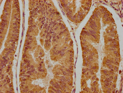
IHC image of CSB-PA856416LA01HU diluted at 1:300 and staining in paraffin-embedded human endometrial cancer performed on a Leica BondTM system. After dewaxing and hydration, antigen retrieval was mediated by high pressure in a citrate buffer (pH 6.0). Section was blocked with 10% normal goat serum 30min at RT. Then primary antibody (1% BSA) was incubated at 4°C overnight. The primary is detected by a biotinylated secondary antibody and visualized using an HRP conjugated SP system.
.")
IHC image of CSB-PA856416LA01HU diluted at 1:300 and staining in paraffin-embedded human endometrial cancer performed on a Leica BondTM system. After dewaxing and hydration, antigen retrieval was mediated by high pressure in a citrate buffer (pH 6.0). Section was blocked with 10% normal goat serum 30min at RT. Then primary antibody (1% BSA) was incubated at 4°C overnight. The primary is detected by a biotinylated secondary antibody and visualized using an HRP conjugated SP system.
NSMAF Antibody
CSB-PA856416LA01HU
ApplicationsImmunoFluorescence, ELISA, ImmunoHistoChemistry
Product group Antibodies
ReactivityHuman
TargetNSMAF
Overview
- SupplierCusabio
- Product NameNSMAF Antibody
- Delivery Days Customer20
- ApplicationsImmunoFluorescence, ELISA, ImmunoHistoChemistry
- CertificationResearch Use Only
- ClonalityPolyclonal
- ConjugateUnconjugated
- Gene ID8439
- Target nameNSMAF
- Target descriptionneutral sphingomyelinase activation associated factor
- Target synonymsFAN, GRAMD5, protein FAN, factor associated with N-SMase activation, factor associated with neutral sphingomyelinase activation, neutral sphingomyelinase (N-SMase) activation associated factor
- HostRabbit
- IsotypeIgG
- Protein IDQ92636
- Protein NameProtein FAN
- Scientific DescriptionCouples the p55 TNF-receptor (TNF-R55 / TNFR1) to neutral sphingomyelinase (N-SMASE). Specifically binds to the N-smase activation domain of TNF-R55. May regulate ceramide production by N-SMASE.
- ReactivityHuman
- Storage Instruction-20°C or -80°C
- UNSPSC41116161
Related products
Product group Antibodies
ApplicationsImmunoPrecipitation, Western Blot, ImmunoCytoChemistry, ImmunoHistoChemistry
ReactivityMouse, Porcine, Rat
TargetNSMAF
- SizePrice
Product group Antibodies
Anti-FAN (internal) Antibody107-10400
ApplicationsWestern Blot, ImmunoHistoChemistry, ImmunoHistoChemistry Paraffin
ReactivityHuman
TargetNSMAF
- SizePrice
Product group Antibodies
FAN antibody [N2C1], InternalGTX103723
ApplicationsWestern Blot, ImmunoHistoChemistry, ImmunoHistoChemistry Paraffin
ReactivityHuman
TargetNSMAF
- SizePrice
Product group Antibodies

NSMAF Antibody (aa176-575)LS-C374793
ApplicationsWestern Blot
ReactivityHuman
TargetNSMAF
- SizePrice

Product group Antibodies
Anti-NSMAF AntibodyHPA023067
ApplicationsImmunoHistoChemistry
ReactivityHuman
TargetNSMAF
- SizePrice
Product group Antibodies

ApplicationsWestern Blot, ImmunoHistoChemistry
ReactivityHuman
TargetNSMAF
- SizePrice

Product group Antibodies
Anti-NSMAF Antibody Picoband(r)A09560-CARRIER-FREE
ApplicationsFlow Cytometry, Western Blot, ELISA
ReactivityHuman, Mouse, Rat
TargetNSMAF
- SizePrice