

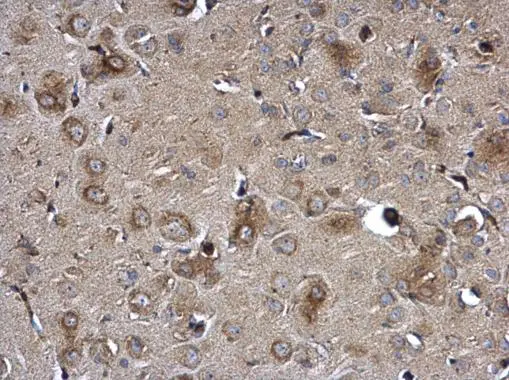
NTE antibody detects NTE protein at cytosol on rat fore brain by immunohistochemical analysis. Sample: Paraffin-embedded rat fore brain. NTE antibody (GTX115501) dilution: 1:500.
Antigen Retrieval: Trilogy? (EDTA based, pH 8.0) buffer, 15min
 dilution: 1:500.
Antigen Retrieval: Trilogy? (EDTA based, pH 8.0) buffer, 15min")
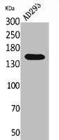
 were separated by 5% SDS-PAGE, and the membrane was blotted with NTE antibody (GTX115501) diluted at 1:1000. The HRP-conjugated anti-rabbit IgG antibody (GTX213110-01) was used to detect the primary antibody, and the signal was developed with Trident ECL plus-Enhanced.")
 were separated by 5% SDS-PAGE, and the membrane was blotted with NTE antibody (GTX115501) diluted at 1:1000. The HRP-conjugated anti-rabbit IgG antibody (GTX213110-01) was used to detect the primary antibody, and the signal was developed with Trident ECL plus-Enhanced. Corresponding RNA expression data for the same cell lines are based on Human Protein Atlas program.")
NTE antibody detects NTE protein at cytosol on rat fore brain by immunohistochemical analysis. Sample: Paraffin-embedded rat fore brain. NTE antibody (GTX115501) dilution: 1:500.
Antigen Retrieval: Trilogy? (EDTA based, pH 8.0) buffer, 15min
NTE antibody
GTX115501
ApplicationsWestern Blot, ImmunoHistoChemistry, ImmunoHistoChemistry Paraffin
Product group Antibodies
ReactivityHuman, Mouse, Rat
TargetPNPLA6
Overview
- SupplierGeneTex
- Product NameNTE antibody
- Delivery Days Customer9
- Application Supplier NoteWB: 1:500-1:3000. IHC-P: 1:100-1:1000. *Optimal dilutions/concentrations should be determined by the researcher.Not tested in other applications.
- ApplicationsWestern Blot, ImmunoHistoChemistry, ImmunoHistoChemistry Paraffin
- CertificationResearch Use Only
- ClonalityPolyclonal
- Concentration0.4 mg/ml
- ConjugateUnconjugated
- Gene ID10908
- Target namePNPLA6
- Target descriptionpatatin like domain 6, lysophospholipase
- Target synonymsBNHS, LNMS, NTE, NTEMND, OMCS, SPG39, iPLA2delta, sws, patatin-like phospholipase domain-containing protein 6, neuropathy target esterase, patatin like phospholipase domain containing 6
- HostRabbit
- IsotypeIgG
- Protein IDQ8IY17
- Protein NamePatatin-like phospholipase domain-containing protein 6
- Scientific DescriptionThis gene encodes a phospholipase that deacetylates intracellular phosphatidylcholine to produce glycerophosphocholine. It is thought to function in neurite outgrowth and process elongation during neuronal differentiation. The protein is anchored to the cytoplasmic face of the endoplasmic reticulum in both neurons and non-neuronal cells. Mutations in this gene result in autosomal recessive spastic paraplegia, and the protein is the target for neurodegeneration induced by organophosphorus compounds and chemical warfare agents. Multiple transcript variants encoding different isoforms have been found for this gene. [provided by RefSeq]
- ReactivityHuman, Mouse, Rat
- Storage Instruction-20°C or -80°C,2°C to 8°C
- UNSPSC41116161
Datasheet
Related products

Product group Antibodies
Anti-PNPLA6 AntibodyA96789
ApplicationsWestern Blot, ELISA
ReactivityHuman, Mouse, Rat
- SizePrice
Product group Antibodies
Anti-PNPLA6 (C-term) Antibody102-22242
ApplicationsWestern Blot
TargetPNPLA6
- SizePrice

Product group Antibodies
Anti-PNPLA6 Antibody Picoband(r)A04071-2-CARRIER-FREE
ApplicationsFlow Cytometry, ImmunoFluorescence, Western Blot, ELISA, ImmunoCytoChemistry
ReactivityHuman
TargetPNPLA6
- SizePrice
Product group Antibodies

PNPLA6 / NTE AntibodyLS-C671712
ApplicationsImmunoFluorescence, ELISA, ImmunoHistoChemistry, ImmunoHistoChemistry Paraffin
ReactivityHuman
TargetPNPLA6
- SizePrice
Product group Antibodies

Goat anti-NTEEB09135
ApplicationsELISA
ReactivityHuman, Mouse
TargetPNPLA6
- SizePrice
Product group Antibodies
PNPLA6 AntibodyCSB-PA005585
ApplicationsWestern Blot, ELISA
ReactivityHuman, Mouse, Rat
TargetPNPLA6
- SizePrice

Product group Antibodies
Anti-PNPLA6 AntibodyHPA007522
ApplicationsImmunoHistoChemistry
ReactivityHuman
TargetPNPLA6
- SizePrice
![Non-transfected (–) and transfected (+) 293T whole cell extracts were separated by 5% SDS-PAGE, and the membrane was blotted with NTE antibody [HL3127] (GTX640613) diluted at 1:50000. The HRP-conjugated anti-rabbit IgG antibody (GTX213110-01) was used to detect the primary antibody.](https://www.genetex.com/upload/website/prouct_img/normal/GTX640613/GTX640613_T-45467_20240913_WB_multiple_B_24091901_979.webp)
Product group Antibodies
NTE antibody [HL3127]GTX640613
ApplicationsWestern Blot
ReactivityHuman, Mouse, Rat
TargetPNPLA6
- SizePrice