

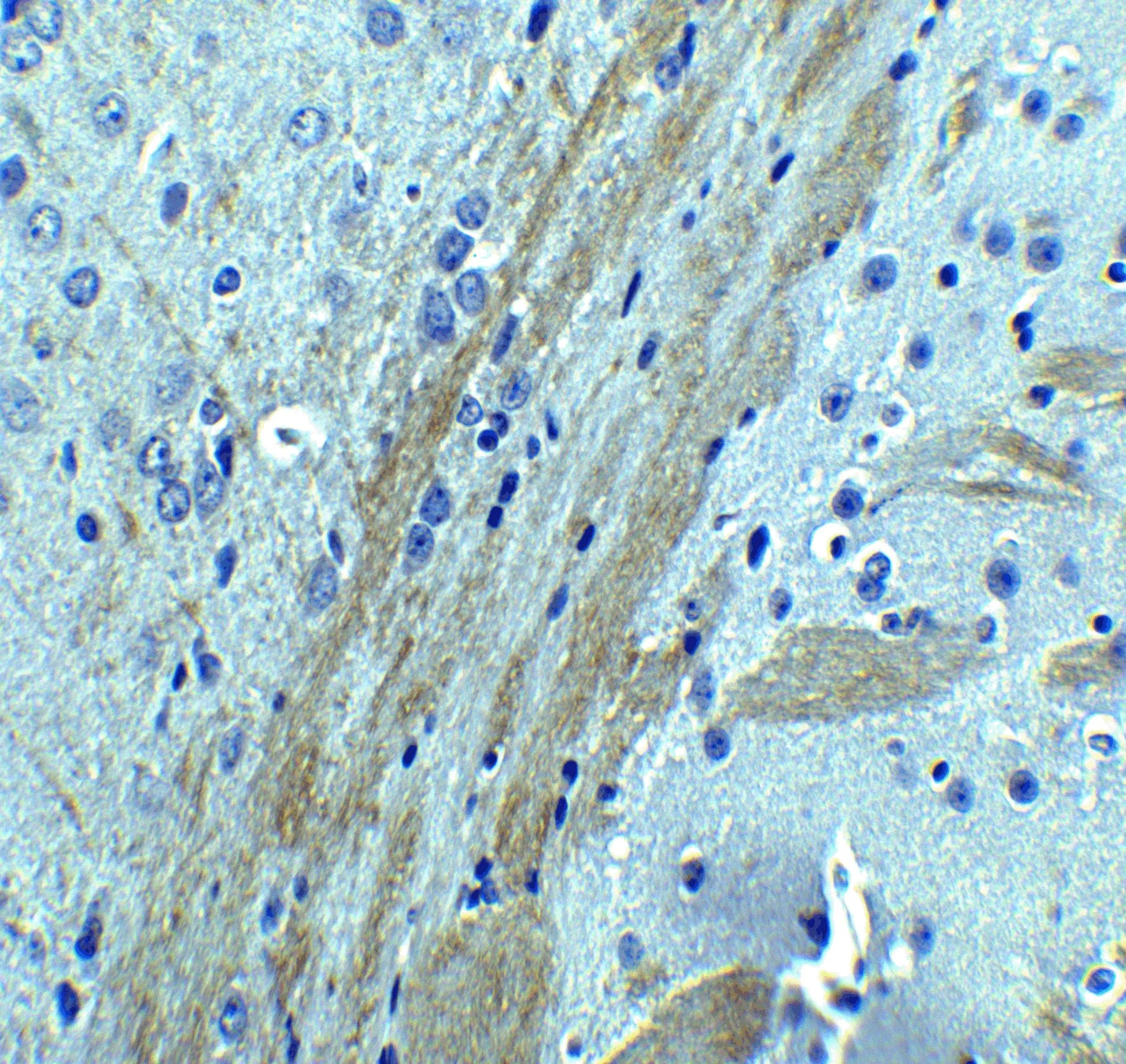
IHC-P analysis of mouse brain tissue using GTX85016 Occludin antibody. Dilution : 5 μg/ml
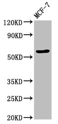
 were separated by 7.5% SDS-PAGE, and the membrane was blotted with Occludin antibody (GTX85016) diluted at 1:500. The HRP-conjugated anti-rabbit IgG antibody (GTX213110-01) was used to detect the primary antibody.")


IHC-P analysis of mouse brain tissue using GTX85016 Occludin antibody. Dilution : 5 μg/ml
Occludin antibody
GTX85016
ApplicationsWestern Blot, ELISA, ImmunoHistoChemistry, ImmunoHistoChemistry Paraffin
Product group Antibodies
ReactivityHuman, Mouse, Rat
TargetOCLN
Overview
- SupplierGeneTex
- Product NameOccludin antibody
- Delivery Days Customer9
- Application Supplier NoteWB: 1 microg/mL. *Optimal dilutions/concentrations should be determined by the researcher.Not tested in other applications.
- ApplicationsWestern Blot, ELISA, ImmunoHistoChemistry, ImmunoHistoChemistry Paraffin
- CertificationResearch Use Only
- ClonalityPolyclonal
- Concentration1 mg/ml
- ConjugateUnconjugated
- Gene ID100506658
- Target nameOCLN
- Target descriptionoccludin
- Target synonymsBLCPMG, PPP1R115, PTORCH1, occludin, phosphatase 1, regulatory subunit 115
- HostRabbit
- IsotypeIgG
- Protein IDQ16625
- Protein NameOccludin
- Scientific DescriptionTight junctions act as a semi-permeable barrier to the transport of ions, solutes, and water and are considered to function as a fence that divides apical and basolateral domains of plasma membranes. Tight junctions coordinate a variety of signaling and trafficking molecules regulating cell differentiation, proliferation, and polarity and contain a number of junctional proteins including Occludin, Claudins, junctional adhesion molecules (JAMs), as well as multiple scaffold proteins. Occludin, the first identified component of tight junction strands, is thought function as a signal transmitter in multiple signaling pathways and can associate with multiple kinases and phosphatases such as PI3-kinase and protein phosphatases 1 and 2A. At least two isoforms of OCLN are known to exist.
- ReactivityHuman, Mouse, Rat
- Storage Instruction2°C to 8°C
- UNSPSC12352203
References
- Wei MF, Cheng CH, Wen SY, et al. Atorvastatin Attenuates Radiotherapy-Induced Intestinal Damage through Activation of Autophagy and Antioxidant Effects. Oxid Med Cell Longev. 2022,2022:7957255. doi: 10.1155/2022/7957255Read this paper
- Li HL, Lu L, Wang XS, et al. Alteration of Gut Microbiota and Inflammatory Cytokine/Chemokine Profiles in 5-Fluorouracil Induced Intestinal Mucositis. Front Cell Infect Microbiol. 2017,7:455. doi: 10.3389/fcimb.2017.00455Read this paper
Datasheet
Related products
Product group Antibodies

Anti-Occludin [1-3]AB04036-10.0
ApplicationsFlow Cytometry, ImmunoPrecipitation, ELISA, ImmunoCytoChemistry, Neutralisation/Blocking, Other Application
ReactivityHuman, Monkey, Mouse, Rat
TargetOCLN
- SizePrice
Product group Antibodies
Anti-OCLN Antibody144-02601
ApplicationsImmunoFluorescence, Western Blot, ImmunoHistoChemistry
ReactivityHuman, Mouse, Rat
TargetOCLN
- SizePrice

Product group Antibodies
Anti-OCLN AntibodyAMAB90889
ApplicationsWestern Blot, ImmunoCytoChemistry, ImmunoHistoChemistry
ReactivityHuman
TargetOCLN
- SizePrice

Product group Antibodies
Anti-Occludin/OCLN Antibody Picoband(r)A01246-2-CARRIER-FREE
ApplicationsFlow Cytometry, ImmunoFluorescence, Western Blot, ELISA, ImmunoCytoChemistry, ImmunoHistoChemistry
ReactivityHuman
TargetOCLN
- SizePrice
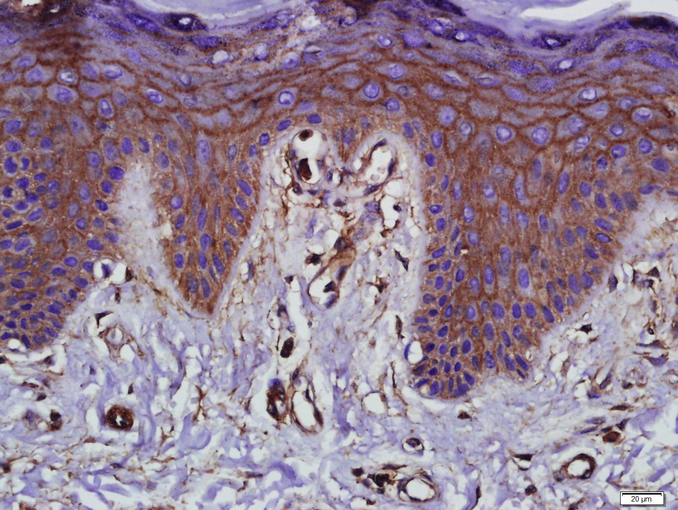
![IHC-P analysis of human ovarian clear cell carcinoma (OCCC) tissue using GTX04461 Occludin antibody [MSVA-415M] HistoMAX?. Clear cell ovarian carcinoma with moderate occludin staining of tumor cells.](https://www.genetex.com/upload/website/prouct_img/normal/GTX04461/GTX04461_20230728_IHC-P_88_23072722_572.webp)
Product group Antibodies
ApplicationsImmunoHistoChemistry, ImmunoHistoChemistry Paraffin
ReactivityHuman
TargetOCLN
- SizePrice
Product group Antibodies
References
Occludin antibodyGTX114949
ApplicationsImmunoFluorescence, Western Blot, ImmunoCytoChemistry, ImmunoHistoChemistry, ImmunoHistoChemistry Frozen, ImmunoHistoChemistry Paraffin
ReactivityHuman, Monkey, Mouse, Rat
TargetOCLN
- SizePrice
Product group Antibodies
Ocln Polyclonal AntibodyCAC11278
ApplicationsImmunoFluorescence, Western Blot, ELISA, ImmunoHistoChemistry
TargetOCLN
- SizePrice
Product group Antibodies
References
Occludin Polyclonal AntibodyBS-10011R
ApplicationsFlow Cytometry, Western Blot
ReactivityBovine, Canine, Equine, Guinea Pig, Human, Mouse, Porcine, Rat
TargetOCLN
- SizePrice
Product group Antibodies
OCLN AntibodyCSB-PA016263LA01HU
ApplicationsImmunoFluorescence, Western Blot, ELISA, ImmunoHistoChemistry
ReactivityHuman
TargetOCLN
- SizePrice