

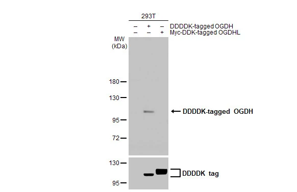
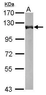
Non-transfected (–) and transfected (+) 293T whole cell extracts (30 μg) were separated by 7.5% SDS-PAGE, and the membrane was blotted with OGDH antibody [HL2072] (GTX637980) diluted at 1:5000. The HRP-conjugated anti-rabbit IgG antibody (GTX213110-01) was used to detect the primary antibody.
![Various whole cell extracts (30 μg) were separated by 7.5% SDS-PAGE, and the membrane was blotted with OGDH antibody [HL2072] (GTX637980) diluted at 1:1000. The HRP-conjugated anti-rabbit IgG antibody (GTX213110-01) was used to detect the primary antibody. Corresponding RNA expression data for the same cell lines are based on Human Protein Atlas program.](https://www.genetex.com/upload/website/prouct_img/normal/GTX637980/GTX637980_T-44893_20240223_WB_TPM_watermark_24022619_584.webp "Various whole cell extracts (30 μg) were separated by 7.5% SDS-PAGE, and the membrane was blotted with OGDH antibody [HL2072] (GTX637980) diluted at 1:1000. The HRP-conjugated anti-rabbit IgG antibody (GTX213110-01) was used to detect the primary antibody. Corresponding RNA expression data for the same cell lines are based on Human Protein Atlas program.")
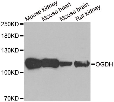
![Various tissue extracts (50 μg) were separated by 7.5% SDS-PAGE, and the membrane was blotted with OGDH antibody [HL2072] (GTX637980) diluted at 1:1000. The HRP-conjugated anti-rabbit IgG antibody (GTX213110-01) was used to detect the primary antibody.](https://www.genetex.com/upload/website/prouct_img/normal/GTX637980/GTX637980_T-44893_20240223_WB_M_tissue_24022619_323.webp "Various tissue extracts (50 μg) were separated by 7.5% SDS-PAGE, and the membrane was blotted with OGDH antibody [HL2072] (GTX637980) diluted at 1:1000. The HRP-conjugated anti-rabbit IgG antibody (GTX213110-01) was used to detect the primary antibody.")
![Rat tissue extract (50 μg) was separated by 7.5% SDS-PAGE, and the membrane was blotted with OGDH antibody [HL2072] (GTX637980) diluted at 1:1000. The HRP-conjugated anti-rabbit IgG antibody (GTX213110-01) was used to detect the primary antibody.](https://www.genetex.com/upload/website/prouct_img/normal/GTX637980/GTX637980_T-44893_20240223_WB_R_muscle_24022619_216.webp "Rat tissue extract (50 μg) was separated by 7.5% SDS-PAGE, and the membrane was blotted with OGDH antibody [HL2072] (GTX637980) diluted at 1:1000. The HRP-conjugated anti-rabbit IgG antibody (GTX213110-01) was used to detect the primary antibody.")
![OGDH antibody [HL2072] detects OGDH protein at mitochondria by immunofluorescent analysis. Sample: HeLa cells were fixed in ice-cold MeOH for 5 min. Green: OGDH stained by OGDH antibody [HL2072] (GTX637980) diluted at 1:500. Red: alpha Tubulin, a cytoskeleton marker, stained by alpha Tubulin antibody [GT114] (GTX628802) diluted at 1:1000. Blue: Fluoroshield with DAPI (GTX30920).](https://www.genetex.com/upload/website/prouct_img/normal/GTX637980/GTX637980_T-44893_20230317_ICC_IF_24030600_347.webp "OGDH antibody [HL2072] detects OGDH protein at mitochondria by immunofluorescent analysis. Sample: HeLa cells were fixed in ice-cold MeOH for 5 min. Green: OGDH stained by OGDH antibody [HL2072] (GTX637980) diluted at 1:500. Red: alpha Tubulin, a cytoskeleton marker, stained by alpha Tubulin antibody [GT114] (GTX628802) diluted at 1:1000. Blue: Fluoroshield with DAPI (GTX30920).")
Non-transfected (–) and transfected (+) 293T whole cell extracts (30 μg) were separated by 7.5% SDS-PAGE, and the membrane was blotted with OGDH antibody [HL2072] (GTX637980) diluted at 1:5000. The HRP-conjugated anti-rabbit IgG antibody (GTX213110-01) was used to detect the primary antibody.
OGDH antibody [HL2072]
GTX637980
ApplicationsImmunoFluorescence, Western Blot, ImmunoCytoChemistry
Product group Antibodies
ReactivityHuman, Mouse, Rat
TargetOGDH
Overview
- SupplierGeneTex
- Product NameOGDH antibody [HL2072]
- Delivery Days Customer9
- Application Supplier NoteWB: 1:500-1:10000. *Optimal dilutions/concentrations should be determined by the researcher.Not tested in other applications.
- ApplicationsImmunoFluorescence, Western Blot, ImmunoCytoChemistry
- CertificationResearch Use Only
- ClonalityMonoclonal
- Clone IDHL2072
- Concentration1 mg/ml
- ConjugateUnconjugated
- Gene ID4967
- Target nameOGDH
- Target descriptionoxoglutarate dehydrogenase
- Target synonymsAKGDH, E1k, E1o, HsOGDH, KGD1, OGDC, OGDH-E1, OGDH2, OGDHD, 2-oxoglutarate dehydrogenase complex component E1, 2-oxoglutarate dehydrogenase, mitochondrial, OGDC-E1, alpha-KGDH-E1, oxoglutarate (alpha-ketoglutarate) dehydrogenase (lipoamide), oxoglutarate decarboxylase, oxoglutarate dehydrogenase (succinyl-transferring), testicular tissue protein Li 131, thiamine diphosphate (ThDP)-dependent 2-oxoglutarate dehydrogenase
- HostRabbit
- IsotypeIgG
- Protein IDQ02218
- Protein Name2-oxoglutarate dehydrogenase complex component E1
- Scientific DescriptionThis gene encodes one subunit of the 2-oxoglutarate dehydrogenase complex. This complex catalyzes the overall conversion of 2-oxoglutarate (alpha-ketoglutarate) to succinyl-CoA and CO(2) during the Krebs cycle. The protein is located in the mitochondrial matrix and uses thiamine pyrophosphate as a cofactor. A congenital deficiency in 2-oxoglutarate dehydrogenase activity is believed to lead to hypotonia, metabolic acidosis, and hyperlactatemia. Alternative splicing results in multiple transcript variants encoding distinct isoforms.[provided by RefSeq, Sep 2009]
- ReactivityHuman, Mouse, Rat
- Storage Instruction-20°C or -80°C,2°C to 8°C
- UNSPSC41116161
Datasheet
Related products
Product group Antibodies
OGDH AntibodyCSB-PA016307LA01HU
ApplicationsELISA, ImmunoHistoChemistry
ReactivityHuman
TargetOGDH
- SizePrice

Product group Antibodies
Anti-OGDH Antibody Picoband(r)A05301-1-CARRIER-FREE
ApplicationsFlow Cytometry, Western Blot, ELISA
ReactivityHuman, Mouse, Rat
TargetOGDH
- SizePrice
Product group Antibodies
Anti-OGDH AntibodyA31346
ApplicationsWestern Blot, ImmunoHistoChemistry
ReactivityHuman, Mouse, Rat
- SizePrice
Product group Antibodies

OGDH AntibodyLS-C747576
ApplicationsImmunoFluorescence, Western Blot, ImmunoHistoChemistry
ReactivityHuman, Mouse, Rat
TargetOGDH
- SizePrice
Product group Antibodies
Anti-OGDH AntibodyHPA019514
ApplicationsImmunoCytoChemistry, ImmunoHistoChemistry
ReactivityHuman
TargetOGDH
- SizePrice
Product group Antibodies
ApplicationsImmunoPrecipitation, Western Blot, ImmunoCytoChemistry, ImmunoHistoChemistry
ReactivityMouse, Rat
TargetOGDH
- SizePrice
Product group Antibodies
OGDH antibody [C2C3], C-termGTX105124
ApplicationsImmunoFluorescence, Western Blot, ImmunoCytoChemistry, ImmunoHistoChemistry, ImmunoHistoChemistry Paraffin
ReactivityHuman, Mouse
TargetOGDH
- SizePrice

Product group Antibodies
References
OGDH antibodyGTX33374
ApplicationsImmunoFluorescence, Western Blot, ImmunoCytoChemistry, ImmunoHistoChemistry, ImmunoHistoChemistry Paraffin
ReactivityHuman, Mouse, Rat
TargetOGDH
- SizePrice