

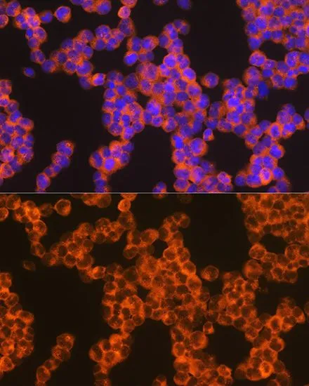
ICC/IF analysis of Jurkat cells using GTX03212 Oncostatin M antibody [GT1300]. Blue : DAPI for nuclear staining Dilution : 1:100
![IHC-P analysis of rat spleen tissue section using GTX03212 Oncostatin M antibody [GT1300]. Blue : DAPI for nuclear staining Dilution : 1:100](https://www.genetex.com/upload/website/prouct_img/normal/GTX03212/GTX03212_20210615_IHC-P_38_w_23053123_358.webp "IHC-P analysis of rat spleen tissue section using GTX03212 Oncostatin M antibody [GT1300]. Blue : DAPI for nuclear staining Dilution : 1:100")
![IHC-P analysis of mouse spleen tissue section using GTX03212 Oncostatin M antibody [GT1300]. Blue : DAPI for nuclear staining Dilution : 1:100](https://www.genetex.com/upload/website/prouct_img/normal/GTX03212/GTX03212_20210615_IHC-P_39_w_23053123_872.webp "IHC-P analysis of mouse spleen tissue section using GTX03212 Oncostatin M antibody [GT1300]. Blue : DAPI for nuclear staining Dilution : 1:100")
![Whole cell extract (30 μg) was separated by 12% SDS-PAGE, and the membrane was blotted with Oncostatin M antibody [GT1300] (GTX03212) diluted at 1:1000. The HRP-conjugated anti-rabbit IgG antibody (GTX213110-01) was used to detect the primary antibody.](https://www.genetex.com/upload/website/prouct_img/normal/GTX03212/GTX03212_4000001194_20210709_WB_2_w_23053123_154.webp "Whole cell extract (30 μg) was separated by 12% SDS-PAGE, and the membrane was blotted with Oncostatin M antibody [GT1300] (GTX03212) diluted at 1:1000. The HRP-conjugated anti-rabbit IgG antibody (GTX213110-01) was used to detect the primary antibody.")
![Various whole cell extracts (30 μg) were separated by 12% SDS-PAGE, and the membrane was blotted with Oncostatin M antibody [GT1300] (GTX03212) diluted at 1:1000. The HRP-conjugated anti-rabbit IgG antibody (GTX213110-01) was used to detect the primary antibody.](https://www.genetex.com/upload/website/prouct_img/normal/GTX03212/GTX03212_4000001194_20210709_WB_w_23053123_983.webp "Various whole cell extracts (30 μg) were separated by 12% SDS-PAGE, and the membrane was blotted with Oncostatin M antibody [GT1300] (GTX03212) diluted at 1:1000. The HRP-conjugated anti-rabbit IgG antibody (GTX213110-01) was used to detect the primary antibody.")
![WB analysis of various samples using GTX03212 Oncostatin M antibody [GT1300]. Dilution : 1:1000 Loading : 25μg per lane](https://www.genetex.com/upload/website/prouct_img/normal/GTX03212/GTX03212_45_WB_w_23053123_167.webp "WB analysis of various samples using GTX03212 Oncostatin M antibody [GT1300]. Dilution : 1:1000 Loading : 25μg per lane")
ICC/IF analysis of Jurkat cells using GTX03212 Oncostatin M antibody [GT1300]. Blue : DAPI for nuclear staining Dilution : 1:100
Oncostatin M antibody [GT1300]
GTX03212
ApplicationsImmunoFluorescence, Western Blot, ImmunoCytoChemistry, ImmunoHistoChemistry, ImmunoHistoChemistry Paraffin
Product group Antibodies
ReactivityHuman, Mouse, Rat
TargetOSM
Overview
- SupplierGeneTex
- Product NameOncostatin M antibody [GT1300]
- Delivery Days Customer9
- Application Supplier NoteWB: 1:500 - 1:2000. ICC/IF: 1:50 - 1:200. *Optimal dilutions/concentrations should be determined by the researcher.Not tested in other applications.
- ApplicationsImmunoFluorescence, Western Blot, ImmunoCytoChemistry, ImmunoHistoChemistry, ImmunoHistoChemistry Paraffin
- CertificationResearch Use Only
- ClonalityMonoclonal
- Clone IDGT1300
- Concentration0.4 mg/ml
- ConjugateUnconjugated
- Gene ID5008
- Target nameOSM
- Target descriptiononcostatin M
- Target synonymsoncostatin-M
- HostRabbit
- IsotypeIgG
- Protein IDP13725
- Protein NameOncostatin-M
- Scientific DescriptionThis gene encodes a member of the leukemia inhibitory factor/oncostatin-M (LIF/OSM) family of proteins. The encoded preproprotein is proteolytically processed to generate the mature protein. This protein is a secreted cytokine and growth regulator that inhibits the proliferation of a number of tumor cell lines. This protein also regulates the production of other cytokines, including interleukin 6, granulocyte-colony stimulating factor and granulocyte-macrophage colony stimulating factor in endothelial cells. This gene and the related gene, leukemia inhibitory factor, also present on chromosome 22, may have resulted from the duplication of a common ancestral gene. Alternative splicing results in multiple transcript variants, at least one of which encodes an isoform that is proteolytically processed. [provided by RefSeq, Jan 2016]
- ReactivityHuman, Mouse, Rat
- Storage Instruction-20°C or -80°C,2°C to 8°C
- UNSPSC12352203
Datasheet
Related products
Product group Antibodies
Anti-OSM Antibody130-10962-500
ApplicationsELISA
ReactivityHuman
TargetOSM
- SizePrice
Product group Antibodies
Osm Polyclonal AntibodyCAC10407
ApplicationsELISA, ImmunoHistoChemistry
TargetOSM
- SizePrice
Product group Antibodies
Oncostatin M Recombinant AntibodyBSM-62322R
ApplicationsWestern Blot
ReactivityHuman, Mouse, Rat
TargetOSM
- SizePrice
Product group Antibodies
Oncostatin M antibody [17001]GTX10842
ApplicationsWestern Blot, ELISA, Neutralisation/Blocking
ReactivityHuman
TargetOSM
- SizePrice
![IHC-P analysis of lung tissue using GTX83959 Oncostatin M antibody [2B6]. Antigen retrieval : Heat-induced epitope retrieval by 10mM citrate buffer, pH6.0, 100oC for 10min. Dilution : 1:50](https://www.genetex.com/upload/website/prouct_img/normal/GTX83959/GTX83959_2050_IHC-P_w_23061420_459.webp)
Product group Antibodies
Oncostatin M antibody [2B6]GTX83959
ApplicationsWestern Blot, ImmunoHistoChemistry, ImmunoHistoChemistry Paraffin
ReactivityHuman
TargetOSM
- SizePrice
Product group Antibodies
Oncostatin M antibody [9G21]GTX52952
ApplicationsWestern Blot, Neutralisation/Blocking
ReactivityHuman
TargetOSM
- SizePrice
Product group Antibodies
OSM AntibodyCSB-PA09694A0RB
ApplicationsELISA, ImmunoHistoChemistry
ReactivityHuman
TargetOSM
- SizePrice
Product group Antibodies

OSM / Oncostatin M AntibodyLS-C403391
ApplicationsELISA, ImmunoHistoChemistry
ReactivityHuman
TargetOSM
- SizePrice